Immune System Glitches. What Causes Long COVID?
Top 24 Potential Causes of Long COVID (LC): Part 1
This disease is not your imagination or your fault!
People with Long COVID experience health problems long after supposedly recovering from COVID-19. If you think you have not completely recovered from COVID-19 you are likely right. Click for a list of some of Long COVID symptoms.
How immune system changes cause Long COVID
The evidence for immune dysfunction in Long COVID is strong. Interestingly, the immune system can crash out either by totally giving up or by becoming hyper alert. One way this may happen is changes to immune cell epigenetics.
Epigenetics refers to how your body can control gene output without changing DNA. Genes express (or produce) the instructions to make different proteins. Epigenetic changes can turn your genes on and off or make genes more or less efficient. Epigenetic markers can change due to environment, nutrition, stress or other factors.
COVID-19 changes immune cell epigenetics
Tran et al. 2024 reported that myeloid and hematopoietic progenitor (parent) cells showed changes in how well the DNA can be accessed (transcriptional and chromatin accessibility changes) after COVID infection. Myeloid and hematopoietic progenitor cells are found in the bone marrow and change into white blood cells (monocytes and granulocytes), red blood cells (erythrocytes), and platelets. This means that there are changes in how genes for white and red blood cells are expressed.
Public Service Announcement:
If you have have chronic fatigue syndrome (CFS), myalgic encephalomyelitis (ME), myalgic encephalomyelitis/chronic fatigue syndrome (ME/CFS) or similar disorders this information may apply to you as well.
Researchers are seeing a lot of similarities between how these these diseases manifest and Long COVID (Komaroff and Lipkin 2023).
Mateo E. (20 year old man): "I was finishing up my junior year of university when I got Long COVID. Truth was I was a bit sluggish but I planned to start cycling. But before I could even start exercising, I caught covid. It's now been 19 months. I'm still enjoying the benefits (ha) of LC like the brain fog, the weird drunk feeling, and fatigue...tired all the freaking time. My 67 year old abuela has more energy than me! I feel like I'm the retiree not her.
I can't work at all right now and get dizzy just standing up. I just want all the chronic illness to go away and to be in better shape. I never wanted to be chronically ill; I wish I could start my life over from scratch and not get COVID. This seems so unfair."
Carl Krenek Sleeping Beauty c1905 tempera.

Part 1 includes six ways your immune system may be sabotaging you after COVID:
1) Unwanted squatter. The virus is still in the house.
2) Your immune cells have zoned out due to Long COVID.
3) Rise of the dead! Virus infection summons zombie COVID viruses hiding in your body.
An additional problem can occur with an overactive immune system; high inflammation levels can trigger and activate glia cells. This can cause chronic pain loops.
5) Self sabotage. Your autoantibodies attack yourself in Long COVID.
6) Too sensitive; the immune system is dialed up too high after COVID.
1) Like an unwanted house guest, the COVID virus is still present and causing problems:
The COVID-19 virus, SARS-CoV-2, lingers in the body even after the person is no longer infectious. These viral reservoirs may contribute to LC since your body is still battling them. Think of the viruses as an entrenched terrorist group fighting a gorilla style war with your bodies's immune cells. They hide in fat cells and organs, put on masks to deceive immune cells, and strike when your immune system is engaged elsewhere. Sneaky viruses have been detected lurking in many organs in the body (discussion in Turner et al. 2023). These dangerous viruses may also be hanging out in your gut causing microbiota dysfunction.
Virus pathogens are associated with other chronic disorders such as myalgic encephalomyelitis (ME) and chronic fatigue syndrome (CFS). Human herpes viruses, B19V, enteroviruses and other viruses can remain latent in the body only to reactivate later and trigger ME and CFS. This is because viral infections can cause changes to host cells which contribute to ME and CFS development. These include chronic inflammation, autoimmunity, immune cell alterations, and mitochondrial modulation (Rasa et al. 2018).
Places that COVID-19 likes to hang out:
Right now it looks like COVID-19 can hide everywhere in the body including the gut (Natarajan et al. 2022), kidney, and spleen (Gupta et al. 2022). Stein et al. 2022 found evidence of the COVID-19 virus in almost all areas of the body including lungs, heart, brain, gut and eyes. Evidence of the virus was found up to 7 months after infection in autopsies of 44 patients with Long Covid.
COVID-19 loves fat cells. SARS-CoV-2, the virus commonly known as COVID-19, can infect and hide out in human fat cells (Martnez-Coln et al. 2022). This may be why obesity is an independent risk factor for Long COVID.
People who are overweight have a 20% increased risk of Long COVID while people who are obese have a 36% increased odds of developing Long COVID. The risk of Long COVID was 20% higher for each 5kg/m2 increase in BMI.
Fat cells can cause other problems like increased inflammation.
Science Bite: SARS-CoV-2 uses metal to hide from immune system
The COVID-19 virus, SARS-CoV-2, uses metal ions; like magnesium, manganese, and calcium; to change the structure of its own RNA and hide from your body's immune system (Viswanathan et al. 2021).
The virus uses metal ions for an architectural purpose; the ions form a bridge between viral messenger RNA (the instructions for encoding the virus) and a protein complex containing viral proteins nsp16 and nsp10. This stabilizes the whole area and allows nsp16 to modify the mRNA cap to make it unrecognizable to our immune system. The virus can than sneak by the immune defences.
Now that is totally metal. Give your body a chance to fight back by getting vaccinated against COVID-19.
2) Your immune cells quietly quit in Long COVID:
Your immune cells are exhausted and communication in the immune system has broken down. It is one big toxic workplace with zoned out employees gossiping and panicking about the big COVID takeover. Long COVID is defined by out of touch immune cells including dysfunctional T cells; CD4+ T cells with increased inflammation; dysregulated immune cells overall; and a lack of crosstalk or coordination between your B and T cells (Yin et al. 2023). It makes The Office look functional.
Learn More:
Here is a quick note on immune cells; CD4 cells fight against infections; CD8 cells kill invaders and cancer cells; and B cells can make antibiotics.
This is an extremely simplified answer. The immune system is so much more complicated than I can cover here. For a quick and fun overview check out this You Tube channel: Kurzgesagt - In a Nutshell How The Immune System ACTUALLY Works IMMUNE. They have a very interesting series on the immune system and an excellent book designed for everyone. Immune: A Journey into the Mysterious System That Keeps You Alive by Phillip Dettmer.
Kurzgesagt - In a Nutshell does not pay me, sadly we don't hang out and play pickleball, and I am not affiliated with them; I just really like their passion for teaching science and philosophy through art.
T cells in Long COVID are really tired of the daily grind. These infection fighting lymphocytes (white blood cells) are still fighting after the ill person is supposedly better. They might be battling a hidden reservoir of SARS-CoV-2 (the COVID-19 virus) or wrestling with a reactivated pathogen like Epstein-Barr virus (EBV), herpes simplex virus, or Hepatitis C (Klein et al. 2022, Davis et al. 2023, discussion in Turner et al. 2023). Yin et al. 2023 found that the cells specifically fighting COVID-19 (SARS-CoV-2-specific CD8+ T cells) are exhausted from fighting.
Whatever enemy they are fighting, these T cells are tired of the battle. Unfortunately, often these weary warriors will just lay down their guns and stop fighting. That is not good for you or your health.
The immune system can check out even after a mild COVID infection. Kratzer et al. 2024 compared 98 people who had not caught COVID with 106 people that had caught COVID and recovered 10 months ago. COVID patients had a large drop in B and T adaptive immune cells. These are the memory cells that learn to recognize viruses in order to kill them quicker. When compared to the healthy controls the COVID patients had reduced absolute granulocyte, monocyte, and lymphocyte cell counts (T, B, and NK cells).
In addition to troops giving up, armies of CD4+ T immune cells start traveling to already inflamed tissues where they cause more inflammation. Yes, instead of helping out like they should, these inflamed and immature acting immune cells go rogue like sailors on shore leave and cause more trouble by releasing inflammatory cytokines near your healthy cells. The chemicals cause more inflammation which attracts more immune cells to perpetuate a giant self reinforcing loop.
When compared to CD4+ T cells in healthy people, CD4+ T cells from people with Long COVID were in a higher inflammatory state (Yin et al. 2023). A small study also found that women with LC have higher amounts of a subgroup activated by cytotoxic T cells. These T cells are associated with gastrointestinal LC symptoms (Su et al. 2022, Yin et al. 2023).
3) COVID-19 infection summons zombie viruses already laying latent in your body.
Yes, dormant (zombie) viruses may basically raise from the dead due to COVID-19 infection (Klein et al. 2022, Davis et al. 2023, discussion in Turner et al. 2023). NEW FEAR UNLOCKED! When your immune response is weakened or challenged dormant viruses may become reactivated and rise up to wreak havoc on your gene expression, protein production, and immune regulation (Proal et al. 2021).
By the way, this is not limited to COVID-19; other viruses can also activate chronic symptoms using this one simple trick.
Reanimation of other latent viruses may occur during the initial COVID-19 infection. Su et al. 2022 conducted a long term study of COVID-19 patients from initial infection to recovery. They reported that 14% of tested patients had reactivation of Epstein-Barr virus (EBV) that causes mononucleosis (mono), and 25% of people had positive SARS-CoV-2 RNAemia (an pneumonia known as acute respiratory syndrome coronavirus 2). These viruses can make people feel sick for months and may contribute to Long COVID.
Treatment with SARS-CoV-2 antivirals may reduce the risk of Long COVID. People randomly assigned to the antiviral molnupiravir during COVID infection had better outcomes at 3 and 6 months (less Long COVID symptoms, better reported health and wellbeing, and improved quality of life) (discussion Al-Aly 2024).
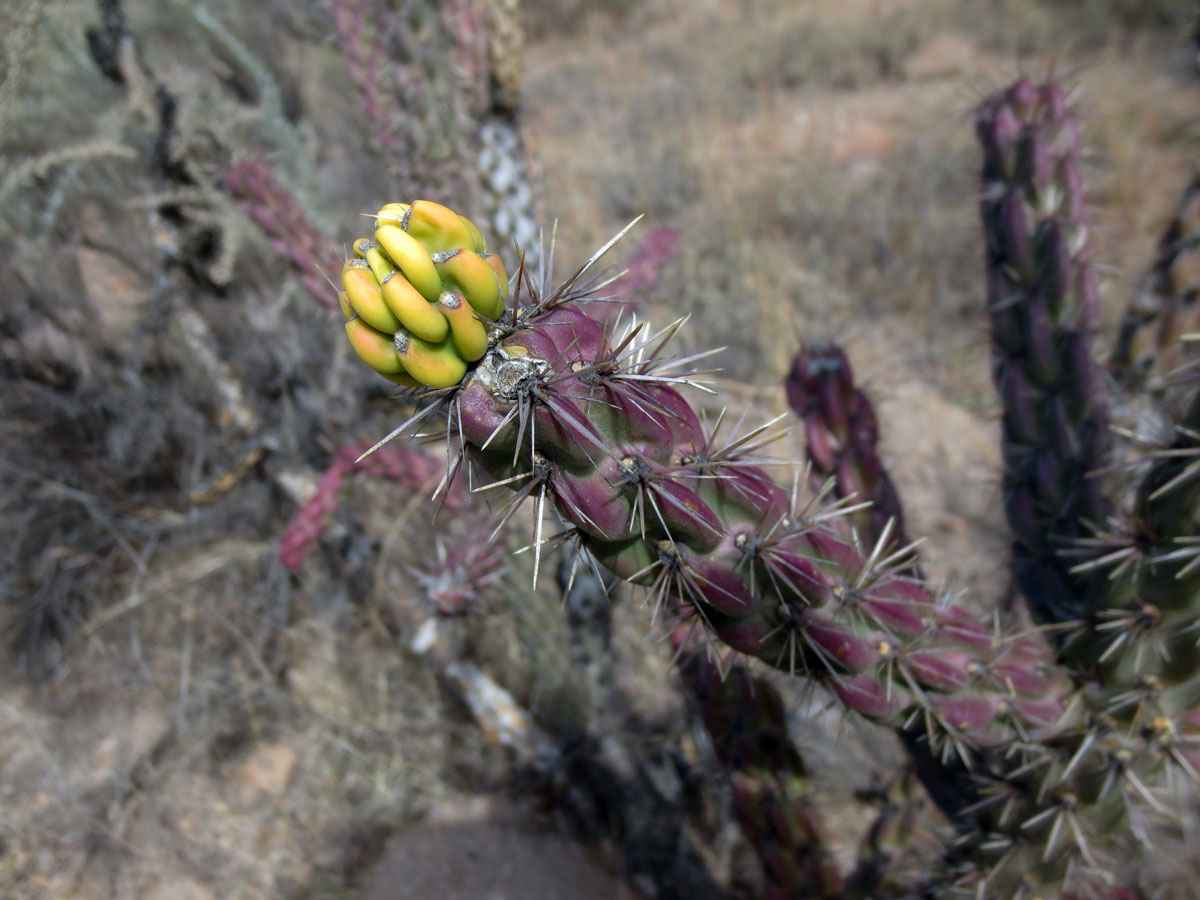
Like a stubborn cactus COVID-19 persists!
4) Instead of quitting; your immune system went full on crazy after COVID infection:
So maybe your immune cells aren't quitters. Maybe they are going all out Rambo style and rooting out every single COVID virus with extreme bloodthirsty violence. What's the problem with such energetic enthusiasm, you may ask?
Unfortunately, once the immune system is activated and destabilized by the COVID-19 virus it may be hard for it to reset to normal. Hypersensitive immune cells attack parts of the body and start to attack your own body in autoimmune reactions. Hyper alertness causes the immune system to go off on normal things that shouldn't be perceived as threats; like your own cells, foods, or pollens.
Indeed, Phetsouphanh et al. 2022, found that people with Long COVID had immune systems that were in a constant state of high alert when compared to people who recovered fully from LC. In Long COVID the white blood cells were highly activated and most of the T cells and B cells had been recruited to fight. In addition to a hyper alert immune systems, people with LC had chronic inflammation.
This is not normal. Healthy people have a large pool of inactive T and B cells just lounging around drinking mock cocktails and chilling. They aren't down to fight all the time; they are just waiting in reserve.
Long COVID also causes dysregulation of the complement system, which is an automatic part of the immune system that contributes to immunity and homeostasis. The complement system is made of a number of distinctly shaped plasma proteins that react and connect with each other to make pathogens easier to kill and induce a series of inflammatory responses that help to fight infection. Think of them as Lego® bricks that can be combined to make all sorts of machinery.
The complement system is part of the innate immune system that targets pathogens and damaged cells (for an amazing video on the complement system see: Tiny Bombs in your Blood - The Complement System by Kurzgesagt in a Nutshell). Watch this video to see these ultra versatile proteins assemble into drilling machines that bore holes in viruses and other pathogens! This kills the virus.
For one year, scientists compared the blood of 113 people with confirmed COVID-19 (SARS-CoV-2) infection with that of 39 people who didn't get COVID. They found that people with Long COVID had changes in blood serum proteins related to the complement immune system.
These changes included activation of the immune system's complement cascade, altered coagulation (blood clotting), and tissue injury such as hemolysis (blood cells bursting), platelets activated and monocyte-platelet clumping (Cervia-Hasler et al. 2024). We are not sure how long it takes for the complement system to recover after COVID-19. Calm down your immune system by frolicking in nature or gazing at your houseplants.
Zara G (29 year old woman): "...before COVID I never got sick - my dad joked around about my epic immune system. I even remember when I caught covid in June 2022. I totally thought my immune system could handle covid and go back to normal, but turns out, no it couldn't.
It got me thinking, some doctors say Long Covid turns into long term autoimmune issues bc the immune system went into warp drive after meeting Covid. It seems that my immune system continued to attack the body even after it killed off the virus. Like it couldn't stop. I was wondering that maybe my immune system is so ridiculously strong that this was why I got long term effects. After I got infected my immune system was clearly ready to kick Covid's butt but unfortunately kept going after Covid was gone and now it doesn't know what to do and is all messed up attacking my body. I have joint pain, chronic fatigue, difficulty breathing and other inflammation problems."
5) After COVID you start making autoantibodies (ANAs) that attack your own body:
As we discussed above, COVID makes your immune system go nuts! After COVID-19 infection, your body can make autoantibodies that attack your own organs and tissues. Viral infections can trigger immune cells' inner Hulk and they start threatening your own cells. New evidence suggest that an autoimmune response plays a role in LC. People with Long COVID have antibodies in their blood that can attack their own tissues. These are called antinuclear/extractable-nuclear antibodies (ANAs).
Compared to healthy people of the same age and gender, people who previously had COVID-19 had high amounts of ANAs in their blood. The ANAs were present in the blood for at least 6-12 months after recovery from COVID (Peker et al. 2021, Lui et al. 2021, Son et al. 2023).
There is a direct connection between high levels of ANAs and inflammation. People with the highest amounts of pro-inflammatory cytokines, such as tumour necrosis factor alpha (TNFα) and C-reactive protein (CRP) had higher ANAs at 1 year after COVID. Likewise, people with the highest levels of TNFa, D-dimer (a product of blood clot breakdown), and interleukin-1β (IL-1β) had worse LC symptoms after 1 year. High concentrations of D-dimer are associated with disease and inflammation. It may also be associated with gut inflammation (Feng et al. 2022).
ANAs are produced when a person's own immune system mistakenly targets and assaults the body's tissues and organs. They are found in people who have chronic inflammation; injury to joints, skin, nervous system, and other tissues; or diseases like lupus or rheumatoid arthritis. If you have chronic inflammation see how inflammation can lead to metabolic disorders. Autoimmune responses like ANAs occur due to exposure to other viruses as well.
The ANAs detected in people with Long COVID even predicted their specific Long COVID syndromes such as fatigue, shortness of breath, and coughing (Son et al. 2023). Men were more susceptible to developing ANAs after COVID-19 infection than women.
U.S. Army Soldiers with the 2nd Battalion, 11th Field Artillery Regiment, 25th Infantry Division fire a M-777A2. U.S. Army photo by Staff Sgt. Ryan Wilhoit c2023.
6) Your immune system is dialed up too high (the interferon pathway) in Long COVID:
Cells release interferon when they sense an infection or invader. Like a warning siren, interferon lets cells know it is time to fight the invading virus. Cells use receptors, such as interferon receptor 2 (IFNA R2), to detect interferon. When infereron signaling is too low or too high it causes the immune system to under or overreact. When your immune system is dialed up too high, you are more likely to get severe or Long COVID.
There are two versions of IFNAR2; a working version and a shorter nonfunctional version. Short IFNAR2 can sense interferon but cannot transmit a signal to cells. It acts as a decoy and interfere with signaling from the normal length IFNAR2. This blunts the cell's immune response to viruses. The ratio of normal to short IFNAR2 may work as a tuning dial to help control the strength of your immune response. People with a abnormally high amount of the short variant of IFNAR2 could be more prone to severe infections; while people with low levels of short IFNAR2 could develop more autoimmune problems like Long COVID (discussion in Pasquesi et al. 2024).
Want more reasons you have Long COVID? Long COVID Part 2: How blood clots, loss of oxygen, nervous system dysfunction and other factors may cause Long COVID.
Definitions:
Brain gut axis is based on how the brain and the gut communication with each other. This complex network involves crosstalk between the vagus nerve; the central nervous system; and gut microbiota; using neural, endocrine, immune, and humoral links. It regulates gastrointestinal homeostasis and it connects the emotional and cognitive areas of the brain with gut functions.
Cytokine storms are life-threatening systemic inflammatory syndromes which involve dangerously high levels of circulating inflammatory chemicals, cytokines, along with hyperactivation of the immune system.
Post-exertional malaise (PEM) which is also called post-exertional symptom exacerbation (PESE) or post-exertional neuroimmune exhaustion(PENE): a medical condition that causes symptoms to get worse after even minor physical or mental exertion. It is common in Long COVID, fibromyalgia, and myalgic encephalomyelitis/chronic fatigue syndrome (ME/CFS).
There is hope for your immune system
After COVID, the immune system resets to normal over 1-2 years
A study looking at 31 women and men with Long COVID (age 40-60) compared to 31 age and sex matched controls found the immune system recovered over time. The abnormally active humoral and cellular immune responses seems to calm down and return to normal after 24 months in most people (Photosphere et al. 2024).
This was a small study but fairly robust. We don't know what happens with the complement system yet.
By Susan Fluegel PHD Nutritional Biochemistry
The immune system is incredibly complex. I took several years of classes in immunity while obtaining my PHD. One of the most important aspects of our immune system is its ability to remember and recognize harmful pathogens and viruses.
Some diseases, such as measles, erase immune memory. That means if you get measles you will wipe out all of your hard built protection against other diseases. To protect yourself and others make sure you are vaccinated against COVID-19 and other diseases. Some people cannot get vaccinated due to their age or health; those of us who can get vaccinated should to help protect the vulnerable.
Konstantin Makovsky An Old Man in Turban (early 1870s).

Some people don't make antibodies when exposed to a disease. They can catch the same disease over and over again. If your diet is low in protein you may not make antibodies when vaccinated. That is why some older people have less immunity to diseases; they have a protein deficiency.
* People are real; names and some other small details have been changed to protect people's privacy.
This information is for informational purposes only and does not constitute medical advice, diagnosis, or treatment.
References:
Al-Aly Z SARS-CoV-2 antivirals and post-COVID-19 condition The Lancet Infectious Diseases, 2024; 25, 6-8 Full article.
Breit S, Kupferberg A, Rogler G, Hasler G. Vagus Nerve as Modulator of the Brain-Gut Axis in Psychiatric and Inflammatory Disorders. Front Psychiatry. 2018 Mar 13;9:44. doi: 10.3389/fpsyt.2018.00044. Full article.
Buoite Stella A, Furlanis G, Frezza NA, Valentinotti R, Ajcevic M, Manganotti P. Autonomic dysfunction in post-COVID patients with and witfhout neurological symptoms: a prospective multidomain observational study. J Neurol. 2022 Feb;269(2):587-596. doi: 10.1007/s00415-021-10735-y. Full article.
Cervia-Hasler C, Brüningk SC, Hoch T, Fan B, Muzio G, Thompson RC, Ceglarek L, Meledin R, Westermann P, Emmenegger M, Taeschler P, Zurbuchen Y, Pons M, Menges D, Ballouz T, Cervia-Hasler S, Adamo S, Merad M, Charney AW, Puhan M, Brodin P, Nilsson J, Aguzzi A, Raeber ME, Messner CB, Beckmann ND, Borgwardt K, Boyman O. Persistent complement dysregulation with signs of thromboinflammation in active Long Covid. Science. 2024 Jan 19;383(6680):eadg7942. doi: 10.1126/science.adg7942. Epub 2024 Jan 19. Full article.
Crow MK, Ronnblom L. Type I interferons in host defence and inflammatory diseases. Lupus Sci Med. 2019 May 28;6(1):e000336. doi: 10.1136/lupus-2019-000336. Full article.
Dai Y, Zhou J, Shi C. Inflammasome: structure, biological functions, and therapeutic targets. MedComm (2020). 2023 Oct 9;4(5):e391. doi: 10.1002/mco2.391. Full article.
de Boer E, Petrache I, Goldstein NM, Olin JT, Keith RC, Modena B, Mohning MP, Yunt ZX, San-Millán I, Swigris JJ. Decreased Fatty Acid Oxidation and Altered Lactate Production during Exercise in Patients with Post-acute COVID-19 Syndrome. Am J Respir Crit Care Med. 2022 Jan 1;205(1):126-129. doi: 10.1164/rccm.202108-1903LE. Full article.
Davis HE, McCorkell L, Vogel JM, Topol EJ. Long COVID: major findings, mechanisms and recommendations. Nat Rev Microbiol. 2023 Mar;21(3):133-146. doi: 10.1038/s41579-022-00846-2. Epub 2023 Jan 13. Erratum in: Nat Rev Microbiol. 2023 Jun;21(6):408. doi: 10.1038/s41579-023-00896-0. Full article.
Excess weight, not high blood sugar, associated with increased risk of COVID-19 infection and long COVID. News release. EurekAlert. September 16, 2022. Accessed September 30, 2022. https://www.eurekalert.org/news-releases/964905
Fernãndez-Castañeda A, Lu P, Geraghty AC, Song E, Lee MH, Wood J, Yalçin B, Taylor KR, Dutton S, Acosta-Alvarez L, Ni L, Contreras-Esquivel D, Gehlhausen JR, Klein J, Lucas C, Mao T, Silva J, Peña-Hernãndez MA, Tabachnikova A, Takahashi T, Tabacof L, Tosto-Mancuso J, Breyman E, Kontorovich A, McCarthy D, Quezado M, Hefti M, Perl D, Folkerth R, Putrino D, Nath A, Iwasaki A, Monje M. Mild respiratory SARS-CoV-2 infection can cause multi-lineage cellular dysregulation and myelin loss in the brain. bioRxiv [Preprint]. 2022 Jan 10:2022.01.07.475453. doi: 10.1101/2022.01.07.475453. Full article.
Feng J, Li J, Li Y, Jin Y, Du F, Chen X. Elevated Serum D-Dimer May Reflect the Presence of Gut Inflammation in Spondyloarthritis. Front Med (Lausanne). 2022 Jan 21;8:816422. doi: 10.3389/fmed.2021.816422. Full article.
Frontera JA, Sabadia S, Yang D, de Havenon A, Yaghi S, Lewis A, Lord AS, Melmed K, Thawani S, Balcer LJ, Wisniewski T, Galetta SL; NYU Neurology COVID-19 Study Team. Life stressors significantly impact long-term outcomes and post-acute symptoms 12-months after COVID-19 hospitalization. J Neurol Sci. 2022 Nov 5;443:120487. doi: 10.1016/j.jns.2022.120487. Full article.
Genecand L, Altarelli M, Binkova A, Loew S, Vaudan S, Gex G, Bridevaux PO, Frésard I. Dysfunctional breathing symptoms, functional impact and quality of life in patients with long COVID-19: a prospective case series. BMJ Open Respir Res. 2023 Jul;10(1):e001770. doi: 10.1136/bmjresp-2023-001770. Full article.
Georgieva E, Ananiev J, Yovchev Y, Arabadzhiev G, Abrashev H, Abrasheva D, Atanasov V, Kostandieva R, Mitev M, Petkova-Parlapanska K, Karamalakova Y, Koleva-Korkelia I, Tsoneva V, Nikolova G. COVID-19 Complications: Oxidative Stress, Inflammation, and Mitochondrial and Endothelial Dysfunction. Int J Mol Sci. 2023 Oct 4;24(19):14876. doi: 10.3390/ijms241914876. Full article.
Giunta S, Giordani C, De Luca M, Olivieri F. Long-COVID-19 autonomic dysfunction: An integrated view in the framework of inflammaging. Mech Ageing Dev. 2024 Apr;218:111915. doi: 10.1016/j.mad.2024.111915. Summary.
Goh D, Lim JCT, Fernaíndez SB, Joseph CR, Edwards SG, Neo ZW, Lee JN, Caballero SG, Lau MC, Yeong JPS. Case report: Persistence of residual antigen and RNA of the SARS-CoV-2 virus in tissues of two patients with long COVID. Front Immunol. 2022 Sep 5;13:939989. doi: 10.3389/fimmu.2022.939989. Erratum in: Front Immunol. 2022 Oct 06;13:1036894. Full article.
Gupta K, Toelzer C, Williamson MK, Shoemark DK, Oliveira ASF, Matthews DA, Almuqrin A, Staufer O, Yadav SKN, Borucu U, Garzoni F, Fitzgerald D, Spatz J, Mulholland AJ, Davidson AD, Schaffitzel C, Berger I. Structural insights in cell-type specific evolution of intra-host diversity by SARS-CoV-2. Nat Commun. 2022 Jan 11;13(1):222. doi: 10.1038/s41467-021-27881-6. Full article.
Greene C, Connolly R, Brennan D, Laffan A, O'Keeffe E, Zaporojan L, O'Callaghan J, Thomson B, Connolly E, Argue R, Meaney JFM, Martin-Loeches I, Long A, Cheallaigh CN, Conlon N, Doherty CP, Campbell M. Blood-brain barrier disruption and sustained systemic inflammation in individuals with long COVID-associated cognitive impairment. Nat Neurosci. 2024 Mar;27(3):421-432. doi: 10.1038/s41593-024-01576-9. Epub 2024 Feb 22. Erratum in: Nat Neurosci. 2024 May;27(5):1019. doi: 10.1038/s41593-024-01644-0. Full article.
Heerdt PM, Shelley B, Singh I. Impaired systemic oxygen extraction long after mild COVID-19: potential perioperative implications. Br J Anaesth. 2022 Mar;128(3):e246-e249. doi: 10.1016/j.bja.2021.12.036. Epub 2021 Dec 27. Erratum in: Br J Anaesth. 2022 Nov;129(5):829-830. Full article.
Kell DB, Laubscher GJ, Pretorius E. A central role for amyloid fibrin microclots in long COVID/PASC: origins and therapeutic implications. Biochem J. 2022 Feb 17;479(4):537-559. doi: 10.1042/BCJ20220016. Full article.
Klein J, Wood J, Jaycox J, Lu P, Dhodapkar RM, Gehlhausen JR, Tabachnikova A, Tabacof L, Malik AA, Kamath K, Greene K, Monteiro VS, Peña-Hernandez M, Mao T, Bhattacharjee B, Takahashi T, Lucas C, Silva J, Mccarthy D, Breyman E, Tosto-Mancuso J, Dai Y, Perotti E, Akduman K, Tzeng TJ, Xu L, Yildirim I, Krumholz HM, Shon J, Medzhitov R, Omer SB, van Dijk D, Ring AM, Putrino D, Iwasaki A. Distinguishing features of Long COVID identified through immune profiling. medRxiv [Preprint]. 2022 Aug 10:2022.08.09.22278592. doi: 10.1101/2022.08.09.22278592. Full article.
Komaroff AL, Lipkin WI. ME/CFS and Long COVID share similar symptoms and biological abnormalities: road map to the literature. Front Med (Lausanne). 2023 Jun 2;10:1187163. doi: 10.3389/fmed.2023.1187163. Full article.
Kratzer B, Gattinger P, Trapin D, et al. Differential decline of SARS-CoV-2-specific antibody levels, innate and adaptive immune cells, and shift of Th1/inflammatory to Th2 serum cytokine levels long after first COVID-19. Allergy. 2024;79:2482-2501. doi:10.1111/all.16210 Full article.
Larsen, Nicholas & Stiles, Lauren & Shaik, Ruba & Schneider, Logan & Muppidi, Srikanth & Tsui, Cheuk & Geng, Linda & Bonilla, Hector & Miglis, Mitchell. (2022). Characterization of autonomic symptom burden in long COVID: A global survey of 2,314 adults. Frontiers in Neurology. 13. 10.3389/fneur.2022.1012668. Full article.
Liu Y, Ebinger JE, Mostafa R, Budde P, Gajewski J, Walker B, Joung S, Wu M, Bräutigam M, Hesping F, Rupieper E, Schubert AS, Zucht HD, Braun J, Melmed GY, Sobhani K, Arditi M, Van Eyk JE, Cheng S, Fert-Bober J. Paradoxical sex-specific patterns of autoantibody response to SARS-CoV-2 infection. J Transl Med. 2021 Dec 30;19(1):524. doi: 10.1186/s12967-021-03184-8. Full article.
Liu Q, Mak JWY, Su Q, Yeoh YK, Lui GC, Ng SSS, Zhang F, Li AYL, Lu W, Hui DS, Chan PK, Chan FKL, Ng SC. Gut microbiota dynamics in a prospective cohort of patients with post-acute COVID-19 syndrome. Gut. 2022 Mar;71(3):544-552. doi: 10.1136/gutjnl-2021-325989. Full article.
Martínez-Colón GJ, Ratnasiri K, Chen H, Jiang S, Zanley E, Rustagi A, Verma R, Chen H, Andrews JR, Mertz KD, Tzankov A, Azagury D, Boyd J, Nolan GP, Schürch CM, Matter MS, Blish CA, McLaughlin TL. SARS-CoV-2 infection drives an inflammatory response in human adipose tissue through infection of adipocytes and macrophages. Sci Transl Med. 2022 Sep 22:eabm9151. doi: 10.1126/scitranslmed.abm9151. Full article.
Molnar T, Lehoczki A, Fekete M, Varnai R, Zavori L, Erdo-Bonyar S, Simon D, Berki T, Csecsei P, Ezer E. Mitochondrial dysfunction in long COVID: mechanisms, consequences, and potential therapeutic approaches. Geroscience. 2024 Apr 26. doi: 10.1007/s11357-024-01165-5. Full article.
Motiejunaite J, Balagny P, Arnoult F, Mangin L, Bancal C, d'Ortho MP, Frija-Masson J. Hyperventilation: A Possible Explanation for Long-Lasting Exercise Intolerance in Mild COVID-19 Survivors? Front Physiol. 2021 Jan 18;11:614590. doi: 10.3389/fphys.2020.614590. Full article.
Natarajan A, Zlitni S, Brooks EF, Vance SE, Dahlen A, Hedlin H, Park RM, Han A, Schmidtke DT, Verma R, Jacobson KB, Parsonnet J, Bonilla HF, Singh U, Pinsky BA, Andrews JR, Jagannathan P, Bhatt AS. Gastrointestinal symptoms and fecal shedding of SARS-CoV-2 RNA suggest prolonged gastrointestinal infection. Med (N Y). 2022 Jun 10;3(6):371-387.e9. doi: 10.1016/j.medj.2022.04.001. Full article.
Norweg A, Yao L, Barbuto S, Nordvig AS, Tarpey T, Collins E, Whiteson J, Sweeney G, Haas F, Leddy J. Exercise intolerance associated with impaired oxygen extraction in patients with long COVID. Respir Physiol Neurobiol. 2023 Jul;313:104062. doi: 10.1016/j.resp.2023.104062. Full article.
Pasquesi GIM, Allen H, Ivancevic A, Barbachano-Guerrero A, Joyner O, Guo K, Simpson DM, Gapin K, Horton I, Nguyen L, Yang Q, Warren CJ, Florea LD, Bitler BG, Santiago ML, Sawyer SL, Chuong EB. Regulation of human interferon signaling by transposon exonization. bioRxiv [Preprint]. 2023 Sep 15:2023.09.11.557241. doi: 10.1101/2023.09.11.557241. Full article.
Peker BO, Sener AG, Kaptan Aydogmus F. Antinuclear antibodies (ANAs) detected by indirect immunofluorescence (IIF) method in acute COVID-19 infection; future roadmap for laboratory diagnosis. J Immunol Methods. 2021 Dec;499:113174. doi: 10.1016/j.jim.2021.113174. Full article.
Phetsouphanh C, Darley DR, Wilson DB, Howe A, Munier CML, Patel SK, Juno JA, Burrell LM, Kent SJ, Dore GJ, Kelleher AD, Matthews GV. Immunological dysfunction persists for 8 months following initial mild-to-moderate SARS-CoV-2 infection. Nat Immunol. 2022 Feb;23(2):210-216. doi: 10.1038/s41590-021-01113-x. Full article.
Phetsouphanh C, Jacka B, Ballouz S, Jackson KJL, Wilson DB, Manandhar B, Klemm V, Tan HX, Wheatley A, Aggarwal A, Akerman A, Milogiannakis V, Starr M, Cunningham P, Turville SG, Kent SJ, Byrne A, Brew BJ, Darley DR, Dore GJ, Kelleher AD, Matthews GV. Improvement of immune dysregulation in individuals with long COVID at 24-months following SARS-CoV-2 infection. Nat Commun. 2024 Apr 17;15(1):3315. doi: 10.1038/s41467-024-47720-8. Full article.
Prasannan N, Heightman M, Hillman T, Wall E, Bell R, Kessler A, Neave L, Doyle A, Devaraj A, Singh D, Dehbi HM, Scully M. Impaired exercise capacity in post-COVID-19 syndrome: the role of VWF-ADAMTS13 axis. Blood Adv. 2022 Jul 12;6(13):4041-4048. doi: 10.1182/bloodadvances.2021006944. Full article.
Proal AD, VanElzakker MB. Long COVID or Post-acute Sequelae of COVID-19 (PASC): An Overview of Biological Factors That May Contribute to Persistent Symptoms. Front Microbiol. 2021 Jun 23;12:698169. doi: 10.3389/fmicb.2021.698169. Full article.
Rasa S, Nora-Krukle Z, Henning N, Eliassen E, Shikova E, Harrer T, Scheibenbogen C, Murovska M, Prusty BK; European Network on ME/CFS (EUROMENE). Chronic viral infections in myalgic encephalomyelitis/chronic fatigue syndrome (ME/CFS). J Transl Med. 2018 Oct 1;16(1):268. doi: 10.1186/s12967-018-1644-y. Full article.
Saito S, Shahbaz S, Luo X, Osman M, Redmond D, Cohen Tervaert JW, Li L, Elahi S. Metabolomic and immune alterations in long COVID patients with chronic fatigue syndrome. Front Immunol. 2024 Jan 18;15:1341843. doi: 10.3389/fimmu.2024.1341843. Full article.
Saleh J, Peyssonnaux C, Singh KK, Edeas M. Mitochondria and microbiota dysfunction in COVID-19 pathogenesis. Mitochondrion. 2020 Sep;54:1-7. doi: 10.1016/j.mito.2020.06.008. Full article.
Singh I, Joseph P, Heerdt PM, Cullinan M, Lutchmansingh DD, Gulati M, Possick JD, Systrom DM, Waxman AB. Persistent Exertional Intolerance After COVID-19: Insights From Invasive Cardiopulmonary Exercise Testing. Chest. 2022 Jan;161(1):54-63. doi: 10.1016/j.chest.2021.08.010. Full article.
Shouman K, Vanichkachorn G, Cheshire WP, Suarez MD, Shelly S, Lamotte GJ, Sandroni P, Benarroch EE, Berini SE, Cutsforth-Gregory JK, Coon EA, Mauermann ML, Low PA, Singer W. Autonomic dysfunction following COVID-19 infection: an early experience. Clin Auton Res. 2021 Jun;31(3):385-394. doi: 10.1007/s10286-021-00803-8. Full article.
Son K, Jamil R, Chowdhury A, Mukherjee M, Venegas C, Miyasaki K, Zhang K, Patel Z, Salter B, Yuen ACY, Lau KS, Cowbrough B, Radford K, Huang C, Kjarsgaard M, Dvorkin-Gheva A, Smith J, Li QZ, Waserman S, Ryerson CJ, Nair P, Ho T, Balakrishnan N, Nazy I, Bowdish DME, Svenningsen S, Carlsten C, Mukherjee M. Circulating anti-nuclear autoantibodies in COVID-19 survivors predict long COVID symptoms. Eur Respir J. 2023 Jan 12;61(1):2200970. doi: 10.1183/13993003.00970-2022. Full article.
Stefano GB, Büttiker P, Weissenberger S, Ptacek R, Wang F, Esch T, Bilfinger TV, Raboch J, Kream RM. Biomedical Perspectives of Acute and Chronic Neurological and Neuropsychiatric Sequelae of COVID-19. Curr Neuropharmacol. 2022;20(6):1229-1240. doi: 10.2174/1570159X20666211223130228. Full article.
Stein SR, Ramelli SC, Grazioli A, Chung JY, Singh M, Yinda CK, Winkler CW, Sun J, Dickey JM, Ylaya K, Ko SH, Platt AP, Burbelo PD, Quezado M, Pittaluga S, Purcell M, Munster VJ, Belinky F, Ramos-Benitez MJ, Boritz EA, Lach IA, Herr DL, Rabin J, Saharia KK, Madathil RJ, Tabatabai A, Soherwardi S, McCurdy MT; NIH COVID-19 Autopsy Consortium; Peterson KE, Cohen JI, de Wit E, Vannella KM, Hewitt SM, Kleiner DE, Chertow DS. SARS-CoV-2 infection and persistence in the human body and brain at autopsy. Nature. 2022 Dec;612(7941):758-763. doi: 10.1038/s41586-022-05542-y. Full article.
Su Y, Yuan D, Chen DG, Ng RH, Wang K, Choi J, Li S, Hong S, Zhang R, Xie J, Kornilov SA, Scherler K, Pavlovitch-Bedzyk AJ, Dong S, Lausted C, Lee I, Fallen S, Dai CL, Baloni P, Smith B, Duvvuri VR, Anderson KG, Li J, Yang F, Duncombe CJ, McCulloch DJ, Rostomily C, Troisch P, Zhou J, Mackay S, DeGottardi Q, May DH, Taniguchi R, Gittelman RM, Klinger M, Snyder TM, Roper R, Wojciechowska G, Murray K, Edmark R, Evans S, Jones L, Zhou Y, Rowen L, Liu R, Chour W, Algren HA, Berrington WR, Wallick JA, Cochran RA, Micikas ME; ISB-Swedish COVID-19 Biobanking Unit; Wrin T, Petropoulos CJ, Cole HR, Fischer TD, Wei W, Hoon DSB, Price ND, Subramanian N, Hill JA, Hadlock J, Magis AT, Ribas A, Lanier LL, Boyd SD, Bluestone JA, Chu H, Hood L, Gottardo R, Greenberg PD, Davis MM, Goldman JD, Heath JR. Multiple early factors anticipate post-acute COVID-19 sequelae. Cell. 2022 Mar 3;185(5):881-895.e20. doi: 10.1016/j.cell.2022.01.014. Full article.
Sureda A, Alizadeh J, Nabavi SF, Berindan-Neagoe I, Cismaru CA, Jeandet P, Los MJ, Clementi E, Nabavi SM, Ghavami S. Endoplasmic reticulum as a potential therapeutic target for covid-19 infection management? Eur J Pharmacol. 2020 Sep 5;882:173288. doi: 10.1016/j.ejphar.2020.173288. Full article.
Tarnawski AS, Ahluwalia A. Endothelial cells and blood vessels are major targets for COVID-19-induced tissue injury and spreading to various organs. World J Gastroenterol. 2022 Jan 21;28(3):275-289. doi: 10.3748/wjg.v28.i3.275. Full article.
Tran BT, Cao R, King KY. Over but not gone: lingering epigenetic effects of COVID-19. Trends Immunol. 2023 Oct;44(10):751-753. doi: 10.1016/j.it.2023.08.012. Full article.
Turner S, Khan MA, Putrino D, Woodcock A, Kell DB, Pretorius E. Long COVID: pathophysiological factors and abnormalities of coagulation. Trends Endocrinol Metab. 2023 Jun;34(6):321-344. doi: 10.1016/j.tem.2023.03.002. Epub 2023 Apr 19. Full article.
Wang S, Quan L, Chavarro JE, Slopen N, Kubzansky LD, Koenen KC, Kang JH, Weisskopf MG, Branch-Elliman W, Roberts AL. Associations of Depression, Anxiety, Worry, Perceived Stress, and Loneliness Prior to Infection With Risk of Post-COVID-19 Conditions. JAMA Psychiatry. 2022 Nov 1;79(11):1081-1091. doi: 10.1001/jamapsychiatry.2022.2640. Erratum in: JAMA Psychiatry. 2022 Sep 28;: Full article.
Wang M, Zhang Y, Li C, Chang W, Zhang L. The relationship between gut microbiota and COVID-19 progression: new insights into immunopathogenesis and treatment. Front Immunol. 2023 May 2;14:1180336. doi: 10.3389/fimmu.2023.1180336. Full article.
Wang B, Zhang L, Wang Y, Dai T, Qin Z, Zhou F, Zhang L. Alterations in microbiota of patients with COVID-19: potential mechanisms and therapeutic interventions. Signal Transduct Target Ther. 2022 Apr 29;7(1):143. doi: 10.1038/s41392-022-00986-0. Full article.
Wong AC, Devason AS, Umana IC, Cox TO, Dohnalová L, Litichevskiy L, Perla J, Lundgren P, Etwebi Z, Izzo LT, Kim J, Tetlak M, Descamps HC, Park SL, Wisser S, McKnight AD, Pardy RD, Kim J, Blank N, Patel S, Thum K, Mason S, Beltra JC, Michieletto MF, Ngiow SF, Miller BM, Liou MJ, Madhu B, Dmitrieva-Posocco O, Huber AS, Hewins P, Petucci C, Chu CP, Baraniecki-Zwil G, Giron LB, Baxter AE, Greenplate AR, Kearns C, Montone K, Litzky LA, Feldman M, Henao-Mejia J, Striepen B, Ramage H, Jurado KA, Wellen KE, O'Doherty U, Abdel-Mohsen M, Landay AL, Keshavarzian A, Henrich TJ, Deeks SG, Peluso MJ, Meyer NJ, Wherry EJ, Abramoff BA, Cherry S, Thaiss CA, Levy M. Serotonin reduction in post-acute sequelae of viral infection. Cell. 2023 Oct 26;186(22):4851-4867.e20. doi: 10.1016/j.cell.2023.09.013. Full article.
Viswanathan T, Misra A, Chan SH, Qi S, Dai N, Arya S, Martinez-Sobrido L, Gupta YK. A metal ion orients SARS-CoV-2 mRNA to ensure accurate 2'-O methylation of its first nucleotide. Nat Commun. 2021 Jun 2;12(1):3287. doi: 10.1038/s41467-021-23594-y. Full article.
Xu R, Lu R, Zhang T, Wu Q, Cai W, Han X, Wan Z, Jin X, Zhang Z, Zhang C. Temporal association between human upper respiratory and gut bacterial microbiomes during the course of COVID-19 in adults. Commun Biol. 2021 Feb 18;4(1):240. doi: 10.1038/s42003-021-01796-w. Full article.
Yin K, Peluso MJ, Luo X, Thomas R, Shin MG, Neidleman J, Andrew A, Young K, Ma T, Hoh R, Anglin K, Huang B, Argueta U, Lopez M, Valdivieso D, Asare K, Deveau TM, Munter SE, Ibrahim R, Ständker L, Lu S, Goldberg SA, Lee SA, Lynch KL, Kelly JD, Martin JN, Münch J, Deeks SG, Henrich TJ, Roan NR. Long COVID manifests with T cell dysregulation, inflammation, and an uncoordinated adaptive immune response to SARS-CoV-2. bioRxiv [Preprint]. 2023 Aug 4:2023.02.09.527892. doi: 10.1101/2023.02.09.527892. Update in: Nat Immunol. 2024 Feb;25(2):218-225. doi: 10.1038/s41590-023-01724-6. Full article.