Exercise Prevents Fat Dysfunction
What is adipocyte (fat cell) dysfunction?
Adipose dysfunction is when fat cells go bad
When fat tissue grows too fast it can become toxic. The fat cells swell up, their lipid (fat) metabolism is impaired, and they start spewing out toxic chemicals (cytokines) which attract immune cells. Immune cells start fights and increase inflammation in the adipose tissue.
Fiberosis develops and blood flow is restricted in the adipose tissue as the inflammation gets worse. This makes it harder for your fat cells to release energy and contributes to insulin resistance. This is why when fat cells go bad you can develop metabolic disorders.
- How exercise helps rescue dysfunctional fat cells
- Your muscles remember your past fitness level
- Gaining fat too quickly messes up your adipose tissue structure causing fibrosis
- What is fibrosis?
- How to treat fibrosis
How exercise rescues dysfunctional adipocytes (fat cells):
1) Exercise decreases lipid (fat) accumulation in fat cells.
Yes, your fat cells can get too fat; this is called adipocyte hypertrophy because it sounds better than GIANT BALLS OF LARD. Hypertrophy (large size) alters how fat cells metabolize lipids and changes the types of adipokines (cytokines) they secretion. Abnormal fat cells produce more inflammatory cytokines that cause inflammation and attract immune cells. Exercise prevents fat gain in fat cells.
Weight gain is the most common cause of fat dysfunction. Somewhere around 75-85% of obese people have dysfunctional adipose tissue. However, not all obese people have dysfunctional extra-large fat cells. People who have normal sized fat cells do not develop impaired adipose tissue. Some skinny people also have toxic dysfunctional fat.
2) Exercise increases antioxidant activity in your body; it increases superoxide dismutase (SOD).
SODs are an important antioxidant defense against oxidative stress. They convert harmful superoxide radicals into harmless chemicals like regular oxygen and water. SODs may inhibit neutrophil-mediated inflammation (Rosa et al. 2021).

3) Exercise decreases insulin resistance while reducing insulin and blood glucose concentrations.
This increases fat cells ability to respond to elevated blood sugar.
4) Exercise increases glucose uptake and insulin sensitivity in white adipose tissue (WAT) (Stroh and Stanford 2023).
5) Exercise increases mitochondria activity in white adipose tissue (Stroh and Stanford 2023).
Mitochondria provide your body with ATP for energy. More active mitochondria mean fat cells are able to burn more fat to make energy.
6) Exercise enhances endocrine activity in white and brown adipose tissue (Stroh and Stanford 2023).
Normal healthy adipose tissue makes and releases hormones that control appetite, insulin sensitivity, inflammation, and various metabolic pathways.
7) Exercise decreases expression of the genes responsible for extracellular matrix (ECM) fibrosis.
Fibrosis is when adipose tissue becomes stiff and scarred making it dysfunctional. Exercise prevents your body from making the proteins; such as collagen, elastin, and fibronectin; needed for fibrosis.
8) Exercise decreases markers of inflammation. This reduces whole body and brain inflammation.
Exercise reduces inflammation by inhibiting the following compounds: NOD-like receptor family pyrin domain-containing 3 inflammasome (NLRP3 inflammasome), nuclear factor-κB (NF-κB), inflammatory cytokines, macrophages, and matrix metalloproteinase-9 (MMP-9).
★ The NLRP3 inflammasome is part of the innate immune system and is normally activated in response to cell damage and microbe invasion (bacterial, fungal, and viral infections). It moderates secretion of proinflammatory cytokines IL-1β and IL-18. High activation rates of the NLRP3 inflammasome is associated with Alzheimer's disease, diabetes, and atherosclerosis (discussion Kelley et al. 2019).
★ NF- κB is a family of transcription factors. Transcription factors are molecules that control gene activity by policing whether DNA gets transcribed into mRNA (mRNA in turn makes proteins). The more a gene is transcribed the more protein it makes. NF- κB is involved in many biological processes, including inflammation, immune responses, cell growth and survival. Too much NF- κB causes an acute inflammation response.
★ Inflammatory cytokines are chemicals that increase inflammation in the body. If you produce too many inflammatory cytokines you develop whole body inflammation.
★ Macrophages are immune cells. Too many immune cells in the wrong place will cause tissue damage (they start firing at everything when excited). Macrophages can infiltrate adipose tissue and cause damage while increasing inflammation.
★ MMP-9, an enzyme involved with remodelling the ECM and maintaining the blood brain barrier. Dysregulated and out of control MMP-9 activity has been associated with tissue destruction, inflammation, neuroinflammation, neurodegenerative diseases, arthritis, cardiovascular diseases, cancer, and autoimmune disorders (discussion Mubarak et al. 2025).
9) Exercise reduces oxidative stress; it lowers reactive oxygen species (ROS) production and hiobarbituric acid reactive substances (TBARS).
Reactive oxygen species (ROS) are important signaling molecules that play a role in the progression of inflammatory disorders and diseases. ROS are used for immune defence and for signaling. However, high levels of ROS cause inflammation.
TBARS is a measure of lipid peroxidation. Lipid peroxidation is when fats degrade due to free radicals and form peroxide and other oxidation products. This is a sign of tissue damage. Fats are a complex nutrient and involved in many biological functions that affect your health.
Discussion in de Sousa Neto et al. 2022, Stroh and Stanford 2023.
Your muscles remember your past fitness level
Your muscles can recall your past levels of fitness. This means out of shape people who used to be fit get in shape faster.
Muscles do this through epigenetics. Epigenetics are markers placed on your DNA that can control the genes. Often a CH3 (methyl group) is either placed on top of the gene or removed.
Genes methylation decreases as you get into shape (hypomethylation). These changes persist even if you revert into being a couch potato (Pilotto et al. 2025).
This means that if you have ever been in shape it will take you less time to get back in shape!
Applied anatomy and kinesiology; the mechanism of muscular movement Photo by Ethel Perrin (1919).
_39_11zon.webp)
Blue Box of Science:
Gaining fat too quickly messes up adipose tissue structure causing fibrosis
Weight gain causes adipose (fat) tissue remodeling. Adipose remodeling is accomplished by the extracellular matrix (ECM). The ECM is a 3-dimensional molecular network that surrounds and supports adipose cells (adipocytes). It is made from collagen I, collagen VII, elastin, fibronectin, glycosaminoglycan (GAG), and laminin. The ECM network serves as mechanical support, maintains normal homeostasisis, and is involved in cell differentiation (when cells decide what they want to be when they grow up), migration, and survival (discussion de Sousa Neto et al. 2022).
The ECM has to change to accommodate the newly created fat cells. Think about gaining weight as trying to stuff more belongings into a full closet. At first the closet shelves are organized and relatively neat. As you randomly toss in spare shoes, extra board games, and that ugly picture of your least favorite cousin (loving gifted from Aunt Maria) the space gets more crowded. The shelves get messier and more disordered. You can't find stuff in the back, air flow is decreased, and everything is pushed together. The same thing happens to your adipose tissue.
Fibrosis is present in metabolically dysfunctional adipose tissue. Adipose tissue expands too rapidly for blood vessels to grow into it. In addition, some of the cytokines produced inhibit angiogenesis (new blood vessel formation). The fat cells start to suffer and die due to lack of oxygen. The cell's lack of oxygen (hypoxia) triggers fibrosis. This becomes a vicious feed back cycle, the stiff ECM in fibrotic fat further impairs adipose tissue blood flow as well as limiting cellular communication.
During fibrosis the ECM gets gummed up due to increased production of ECM proteins such as collagen, elastin, and fibronectin. These proteins cause adipose tissues to thicken and scar. It is similar to the scarring caused by injuries. In fact, fibrosis can also occur due to an injury. Changes in metabolism cause an inflammatory cascade that initiates the fibrotic repair process. Fibrosis is associated with insulin sensitivity, obesity, metabolic dysfunction, and weight loss difficulty (Sun et al. 2013, DeBari and Abbott 2020, de Sousa Neto et al. 2022, Gliniak et al. 2023).
Yes, this means it is harder for people to lose weight once they gain a certain amount of fat (for many people it is when you are around 15% over fat).
Do you want to know more about how fat dysfunction screws up your health?
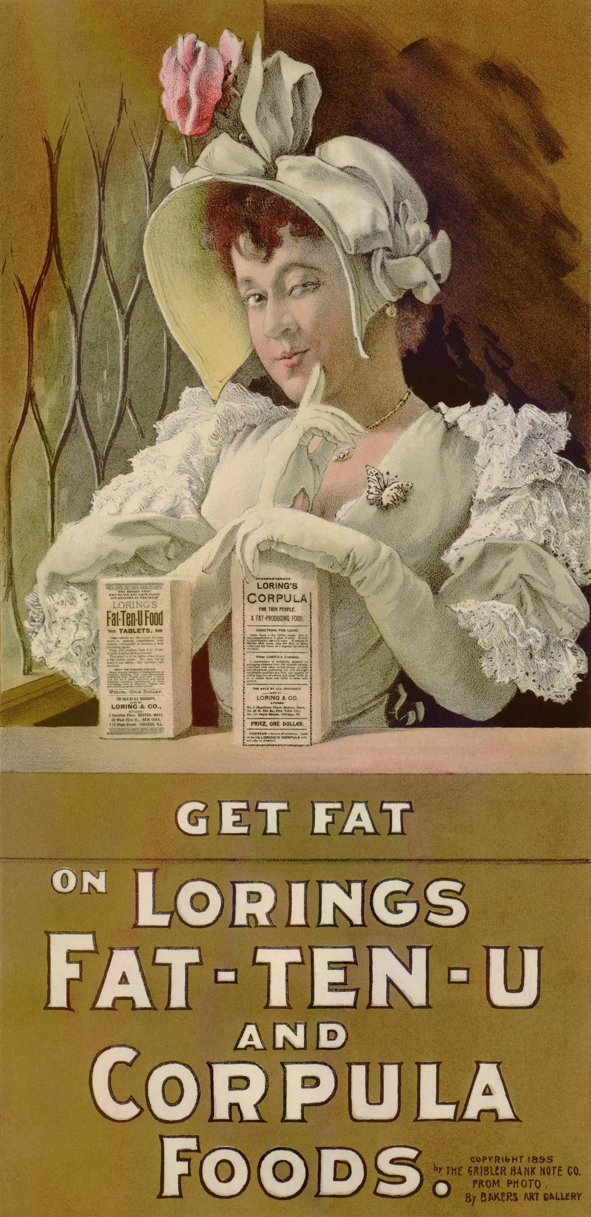
Advertisement showing young woman with package of Loring's Fat-Ten-U from 1895. Fat-Ten-U was a supplement that if taken three times a day would supposedly "produce from 8 to 16 lbs of solid healthy flesh per month."
What is fibrosis?
Fibrosis can also be present in lipedema
During fibrosis the body:
1) Deposits too much fibrous connective tissue, such as collagen, elastin, and fibronectin, in the adipose tissue. This cause it to become scarred and stiff.
2) This lowers blood flow, prevents oxygen from getting into area, and creates inflammation.
3) All of this causes or contributes to metabolic dysfunction including insulin resistance.
4) One end result is that fat cells cannot be easily accessed to use as fuel. This makes weight loss harder.
Signs you have fibrosis:
- Fat feels dense not fluffy.
- You may see small and large bumps in adipose tissue rich areas like butt and thighs.
- It is hard to pinch fat since it is stiff.
- Fatty and lumpy areas feel painful when you touch or gently message them.
*Lisa G. (39 year old woman) "The fibrosis texture ranges. Some tiny bumps are around the size of peas and larger ones feel and look like pebbles. There are some lumps on on my upper and mid thighs that feel like short tubes. Some of the bumps are extremely painful and knotty."
Horse from Santa Fe NM Cathedral Monument.
Burpee's farm annual from 1882. At that time pigs were bred to be very fat.
 YOKKSHIRE PIGS_5_11zon.jpg)
How to treat fibrosis:
Reduce adipose inflammation through anti-inflammatory diet, exercise, and better mental health (which lowers overall inflammation). It may sound funny, but check if your brain needs to heal. Brain inflammation can influence your body inflammation.
Replace inflammatory fats in the diet with better fats. Your body is made of the fats you eat. Seriously, you can do a biopsy of your body fat and count what fats you eat (some fats can be converted to other types but many are used as they come in to the body). If you eat healthier fats it can lower overall inflammation.
Massage and knead areas to increase blood and fluid flow. Consider purchasing a massage unit that mimics kneading to spare your hands. Some units have little balls that roll around and are very effective. Massage and/or vibration plate exercise machines can really help some people.
Tony B. (24 year old woman) "I have fibrosis. I am trying to lower my whole body inflammation by changing my diet. Basically I eliminated sweeteners and processed foods and instead eat tons of vegetables and fruits with some lean meat and fish. And I am seeing an amazing change. Even though I have not lost weight my thighs are skinnier and less bumpy. They feel less painful when I squeeze the fat tissue. It took a couple of months before I started seeing results. I don't plan to go back to my previous diet."
By Susan Fluegel PHD Nutritional Biochemistry and Lori Wood MS Human Nutrition.
*Names and some minor identifying details in all stories in this website are changed to protect people's privacy.
I'm not your doctor; I'm just a person who would like to see you happy and healthy. If you have any questions or doubts about starting an exercise or diet program please consult a professional.
References:
DeBari MK, Abbott RD. Adipose Tissue Fibrosis: Mechanisms, Models, and Importance. Int J Mol Sci. 2020 Aug 21;21(17):6030. doi: 10.3390/ijms21176030. Full article.
de Sousa Neto IV, Durigan JLQ, da Silva ASR, de Cássia Marqueti R. Adipose Tissue Extracellular Matrix Remodeling in Response to Dietary Patterns and Exercise: Molecular Landscape, Mechanistic Insights, and Therapeutic Approaches. Biology (Basel). 2022 May 17;11(5):765. doi: 10.3390/biology11050765. Full article.
Gliniak CM, Pedersen L, Scherer PE. Adipose tissue fibrosis: the unwanted houseguest invited by obesity. J Endocrinol. 2023 Oct 19;259(3):e230180. doi: 10.1530/JOE-23-0180. Full article.
Kelley N, Jeltema D, Duan Y, He Y. The NLRP3 Inflammasome: An Overview of Mechanisms of Activation and Regulation. Int J Mol Sci. 2019 Jul 6;20(13):3328. doi: 10.3390/ijms20133328. Full article.
Pilotto AM, Turner DC, Mazzolari R, Crea E, Brocca L, Pellegrino MA, Miotti D, Bottinelli R, Sharples AP, Porcelli S. Human skeletal muscle possesses an epigenetic memory of high-intensity interval training. Am J Physiol Cell Physiol. 2025 Jan 1;328(1):C258-C272. doi: 10.1152/ajpcell.00423.2024. Full article.
Mubarak MM, Baba IA, Wani ZA, Kantroo HA, Ahmad Z. Matrix metalloproteinase-9 (MMP-9): A macromolecular mediator in CNS infections: A review. Int J Biol Macromol. 2025 Jun;311(Pt 2):143902. doi: 10.1016/j.ijbiomac.2025.143902. Full articles.
Stroh AM, Stanford KI. Exercise-induced regulation of adipose tissue. Curr Opin Genet Dev. 2023 Aug;81:102058. doi: 10.1016/j.gde.2023.102058. Full article.
Sun K, Tordjman J, Clément K, Scherer PE. Fibrosis and adipose tissue dysfunction. Cell Metab. 2013 Oct 1;18(4):470-7. doi: 10.1016/j.cmet.2013.06.016. Full article.
Rosa AC, Corsi D, Cavi N, Bruni N, Dosio F. Superoxide Dismutase Administration: A Review of Proposed Human Uses. Molecules. 2021 Mar 25;26(7):1844. doi: 10.3390/molecules26071844. Full article.