Beyond the Immune System. What Causes Long COVID?
Top 24 Potential Causes of Long COVID (LC): Part 2
Viral infections can gut your body in ways that don't include the immune system. This includes attacks on your nervous system, gut bacteria, oxygen supply, blood and more.
There are many ways that a post viral infection can still influence your health.
Long COVID causes likely apply to other post viral infections such as chronic fatigue syndrome (CFS), myalgic encephalomyelitis (ME), myalgic encephalomyelitis/chronic fatigue syndrome (ME/CFS) or similar disorders.
Read below for nine scary ways COVID-19 can wreck your heath:
- 7) COVID causes abnormal blood clotting.
- 8) COVID viral infection changes how RNA is built in host cells.
- 9) Long COVID drains your energy due to metabolomic and immune disturbances.
- 10) COVID-19 cuts off your oxygen supply.
- 11) Don't freak out about this though; stress makes getting Long COVID worse.
- 12) COVID decreases cortisol causing weakness and depression.
- 13) The COVID virus causes nervous system dysfunction.
- 14) COVID hammers your gut bacteria.
- 15) Your hypothalamus-pituitary-adrenal axis (HPA axis) is disrupted by COVID.
- 16) Endothelial dysfunction drives oxidative stress and inflammation in Long COVID.
Lee Russell 1937 Exhausted flood refugee resting, Sikeston, Missouri.

If you cannot exercise you may have abnormal blood clotting:
People with Long COVID and exercise intolerance had a higher chance of abnormal blood clotting. Those with raised levels of blood clotting markers were 4X more likely to have an impaired exercise capacity.
Researchers measured exercise intolerance by having people with Long COVID perform timed exercises like walking on a treadmill. They tested oxygen levels using a finger sensor as well as blood before and after the exercise. People with exercise intolerance used oxygen up more rapidly while working out and produced more lactate acid than normal (lactate acid is normally produced during exercise).
Researchers also measured abnormal blood clotting markers by looking at the relative levels of two proteins in the body; Von Willebrand factor (VWF), which is a blood clotting protein, and ADAMTS13, a protein that regulates VWF by cutting or splicing it to prevent it from clotting in blood vessels. You need these two proteins to be in balance for normal blood clotting.
People with Long COVID were more likely to have too much VWF; this would cause abnormal blood clotting (Presentee et al. 2022).
7) Long COVID causes abnormal blood clotting full of strange microscopic structures:
Long COVID is associated with microclots; small weird blood clotting proteins. Long COVID sufferers had significantly more and larger microclots (Thierry et al. 2025).
Microclots clump together due to sticky immune webs called neutrophil extracellular traps, or NETs. These NETs may stabilize the microclots and prevent them from being dissolved.
The innate immune response known as NETosis, normally produces NETs. Like Spiderman, neutrophils eject their DNA to form thread-like webs (filamentous). The webs are embedded with cytotoxic enzymes to quickly trap and neutralize pathogens and other dangers to the body. This can go horribly wrong though.
Why are microclots so bad:
Abnormal microclots form dense matted clots that are fibrinolytic‐resistant. This means they are hard for your body to dissolve. These tangled microclots entrap different inflammatory molecules (which include compounds that prevent clot breakdown). All of these proteins combine in one big messy clump.
The mere presence of SARS‐CoV‐2 S1 spike protein causes fibrinolytic ‐resistant microclots to form (discussion in Thierry et al. 2025). Lung biopsies from people who died of COVID-19 showed excess NETs formation with neutrophilic infiltration (Collete et al. 2025).
Excess NETs formation may contribute inflammatory and blood clotting diseases, including autoimmune disorders, pulmonary embolism, deep vein thrombosis, severe infections, cancer, diabetes, and arthritis (discussion in Thierry et al. 2025).
8) COVID-19 infection changes how RNA is built:
Infection by SARS-CoV-2 causes changes in gene expression and transcript usage.
Gene expression is the process by which information copied from DNA is used to make a functional product; usually a protein or RNA. Transcription is when an RNA copy is made of a DNA gene segment. The RNA copy is used to carry the gene's instructions to ribosomes. Ribosomes make proteins using these instructions.
When COVID-19 strikes it can change gene expression influencing blood coagulation, neutrophil activation, and cytokine production. The COVID-19 virus changes the original splicing patterns of numerous cellular transcripts. More severe cases of COVID-19 had greater transcript dysregulation.
The virus often targets the cellular spliceosome. The cellular spliceosome is an extremely large and complex multi-megadalton (megadalton has a mass equal to one million daltons) ribonucleoprotein (RNP) complex (Wang et al. 2022).
9) Long COVID drains your energy due to metabolomic and immune disturbances:
Just like the COVID pandemic did in real life, Long COVID disrupts the pathways and supply chains in your body that make energy and build infrastructure. We can see this disruption by studying the chemicals created by these metabolic pathways; this is called metabolomics.
Metabolomics are the study of small molecules, called metabolites, that are within tissues, cells and fluids of organisms. Metabolites are created when the body runs chemical reactions during metabolizing. They are used for energy, as building blocks, to create structure, as fuel, for cell signalling, as cofactors or catalysts for enzymes, for defence, in epigenetics, during interactions with other organisms and for other purposes. The human metabolic database had 217,920 compounds (metabolites) in 2022 (Wishart et al 2022).
Filippo Palizzi - Pastore addormentato con il suo cane (1850-1855).
_53_11zon.jpg)
People with COVID-19, people with Long COVID, people who recovered from COVID-19 and appeared healthy, and healthy people all show different metabolome profiles. In addition, many metabolic pathways are altered due to catching COVID-19, recovering from the virus or living with LC (Saito et al. 2024).
Saito et al. 2024 main findings:
People with Long COVID (LC) had less plasma ATP (energy).
★ People who currently are ill with an acute COVID-19 infection had 29 metabolic pathways that were significantly altered compared to healthy people.
★ People with LC had 18 metabolic pathways that were significantly different from healthy people AND people who had recovered from LC (15 of these pathways were the same):
∇ The changes in the metabolic pathways meant the levels of aspartate (amino acid- AA), uracil, serine (AA), sarcosine (AA), arginine (AA), dehydroalanine, thymine, and porphobilinogen were lower in LC patients than healthy people. To understand the impact of this: Thymine is one of the bases used in building DNA while uracil is used to make RNA. Porphobilinogen helps build hemoglobin in red blood cells. Amino acids build proteins. This means that people with LC have less of the building blocks needed to make DNA, RNA, proteins and red blood cells.
∇ The levels of other metabolites; such as 5-Aminolevulinate, cysteate, putrescine, 4-Aminobutyraldehyde, kynurenine, serotonin, Formyl-5-hydroxykynurenamine, 5-Hydroxykynurenine, 2-Aminomuconate, xanthine, and 5-Aminolevulinate; were higher in people with LC compared to healthy people.
★ People who had seemly recovered from COVID had alterations in 15 metabolomics pathways when compared to healthy people. Of these, 13 pathways were similar to those found in LC. The other two were vitamin B6 and glutathione metabolisms.
★ Amino acid and nitrogen metabolism was disturbed in many cases.
★ People with LC had a significant reduction in two amino acids; sarcosine, found in muscles, and serine, used to make proteins, nucleic acids, antibiotics, and folate. Sarcosine and serine amounts were inversely correlated with depression, anxiety, and cognitive dysfunction scores. In other words, decreased levels of sarcosine and serine were associated with increased anxiety, depression and cognitive dysfunction.
★ People with Long COVID had elevated proinflammatory cytokines such as IL-1α, IL-6, TNF-α, Flt-1, and sCD14 when compared to people who were healthy or who had recovered from COVID. This means those with LC had a higher level of inflammation. If you have PTSD or cPTSD, you may need to heal to reduce inflammation.
Check out the cool charts in Saito et al. 2024 for all the details!
10) Long COVID cuts off your oxygen:
COVID-19 patients hyperventilate when exercising. Hyperventilation is breathing deeper and faster than normal. It increases the amount of carbon dioxide in the body and can cause light-headedness, muscle spasms, rapid heartbeat, shortness of breath, numbness and chest pain.
Hyperventilation in Long COVID may be due to irregularities in the physiological mechanisms involved in the control of breathing (called ventilatory control) (Mutagenic et al. 2021).
Problems with breathing may contribute to low oxygen uptake. Genevans et al. 2023 found that 1/3 of 264 people with Long COVID had dysfunctional breathing. People in this group (48 total patients) reported that they had had hyperventilation (21%), periodic deep sighs/erratic breathing (47%), and mixed types of dysfunctional breathing (33%) (Genevans et al. 2023). Almost all of them also had fatigue, difficulty focusing, muscle aches and pains (myalgia), forgetfulness and trouble sleeping.
Several small studies suggested that during intense exercise people with Long COVID may have impaired tissue oxygen consumption and oxygen extraction when compared to age matched healthy people (Singh et al. 2022, Heerdt et al. 2022). They found that people who had recovered from COVID infection had reduced peak exercise aerobic capacity (oxygen consumption [VO2]) when compared with control participants. VO2, measures the amount of oxygen a person's body uses during intense exercise. It is usually measured in milliliters of oxygen consumed per minute per kilogram of body weight (mL/kg/min).
This reduction in peak VO2 was connected with impaired systemic oxygen extraction. The muscle cells of people with Long COVID cannot take in oxygen. LC patients with exercise intolerance, may have a skeletal muscle myopathic process (muscle disease) that causes hypoxia (Sing et al. 2022, Norweg et al. 2023). Hypoxia is when your tissues and muscles do not take up oxygen. Basically, blood delivers the oxygen but the tissues can not take it up.
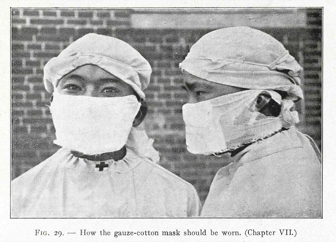
How the gauze-cotton mask should be worn c1910s authors K. Chimin Wong (王吉民) and Wu Lien-Teh (伍連德).
Basically, people who had COVID previously are using less oxygen during exercise and their tissues are having a harder time removing oxygen from the blood. Some symptoms of Long COVID; such as fatigue, post-exertional malaise, and dyspnea; may be due to dysfunctional oxygen uptake, delivery, and ventilation.
 11) Long COVID is more common if you have experienced a stressful event or you are in psychological distress:
If you experience the death of a loved one, financial troubles, food insecurity, or a new developed disability you are twice as likely to have symptoms of Long COVID a year after recovery from COVID-19 (Frontera et al. 2022). Likewise, being in psychological distress such as depression, anxiety, worry, perceived stress, and/or loneliness, before catching COVID-19 was associated with up to a 45% increase in your chances of developing Long COVID (Wang et al. 2022).
Well, that stinks...
If you have depression or anxiety see if you can get some help. Since not everyone has access to professional mental health care or the money to buy it we include some information on how to fight back against depression.
You are important and worthy of being kind to yourself. You matter enough that we have created this website to help YOU (it certainly wasn't for financial gain ha, ha). Take care of yourself. If you feel suicidal please call or text a help line (some contacts here); if you know someone who feels suicidal here is what you can do to try to help prevent suicide.
12) Low cortisol causes weakness and depression in Long COVID:
People with LC often have low concentrations of cortisol (Klein et al. 2022, Giunta et al. 2024). Cortisol is a stress hormone that effects almost every system in your body. It helps regulate inflammation; fine tunes the use of fats, proteins and glucose in the body; controls your wake/sleep cycles; controls motivation, mood and fear; and regulates blood pressure.
We all hear that high cortisol concentrations are bad but low cortisol is also harmful. Too little cortisol can cause muscle weakness, low blood glucose, loss of appetite, nausea, low blood pressure, extreme fatigue, muscle and joint pain, depression, irritability and hair loss. One cause of high cortisol is exposure to abuse and/or trauma which can result in PTSD and cPTSD.
If your childhood trauma was the result of being indoctrinated into a church or cult you may want to read about how to recover from religious trauma. Many churches use guilt and negativity to keep their members compliant.

Low cortisol may be due to hormones being out of balance. See 15) Your hypothalamus-pituitary-adrenal axis (HPA axis) is disrupted.
13) You may have an Autonomic Nervous System (ANS) dysfunction with Long COVID:
The autonomic nervous system (ANS) regulates body processes; like blood pressure, blood clotting, digestion, and breathing; that work automatically (autonomously), without you having to think about it. It helps regulate inflammation, cognition, immune function, blood clotting (coagulation) pathways, fatigue, cognition, exercise intolerance, and other factors that may contribute to Long COVID.
The ANS has three parts:
1) Sympathetic nervous system: activates when it thinks you are in danger or stressed. It is responsible for your body's fight, flight, freeze response. You can help regulate your sympathetic nervous system by exposing yourself to green plants and nature.
2) Parasympathetic nervous system: counteracts the sympathetic nervous system by promoting your ability to relax. Maintains resting heart rate, metabolism and breathing rates.
3) Enteric nervous system: a network of nerves associated with the gastric tract that can act independently of the other two systems. Some people think it is part of the nervous system. It manages how your body digests food by regulating gut secretions, water and electrolytes.
Researchers used the composite autonomic symptom scale 31 (COMPASS 31) questionnaire to help determine the health of the autonomic nervous system in 2,314 women and men (87% were women between 31-65 years old). They looked at 53 different symptoms.
COMPASS 31 is a self-rating questionnaire that helps evaluate six areas of autonomic function: orthostatic intolerance (your ability to remain upright), vasomotor (body temperature dysfunction like hot flashes), secretomotor (ability of glands, like sweat glands, to secrete a substance), gastrointestinal (digestion), bladder (integration between the autonomic and somatic nervous systems), and pupillomotor (controls changes in the eye pupil). COMPASS-31 scores are added over the six autonomic systems to a total score of 1-100; a higher score is more severe (Shouman et al. 2021).
A COMPASS-31 score of above 20 was found in 67% of people with Long COVID. This score shows that the autonomic dysfunction is moderate to severe (Larsen et al. 2022). Most people with Long COVID have a autonomic system that is not working properly.
The severity of COVID-19 did not match with the severity of the autonomic dysfunction; even very mild SARS-CoV-2 infections can cause major autonomic dysfunction (Larsen et al. 2022).
14) Your gut and respiratory microbiota are altered in Long COVID:
The gut and respiratory microbiota are significantly altered in patients with active COVID-19 and Long COVID (Xu et al. 2021, Wang et al. 2022). Microbiota, the community of micro-organisms that live in your gut, are powerful immunomodulatory factors in different human diseases, including cancer, type 2 diabetes, obesity, ulcerative colitis, Crohn’s disease, and some viral infections.
Changes in gut bacteria, called gut dysbiosis, is associated with Long COVID. Six months after recovery from COVID-19, the gut microbiota of patients without Long COVID were similar to healthy people. People with Long COVID, however, had substantially different gut micro-organism communities from those of the healthy people. Notably, people with Long COVID had reduced bacteria diversity and richness when compared to the healthy individuals (Lui et al 2022, Wang et al. 2023). The bacteria population contained more conditional pathogens and opportunistic microorganisms.
This can have an effect on the immune system. For a more complete list of depleted and enriched microbe species due to COVID-19 see Table 1 and 2 in Wang et al. 2023. There is some evidence that full recovery from COVID may restore gut microbiota (discussion in Wang et al. 2023).
15) Your hypothalamus-pituitary-adrenal axis (HPA axis) is disrupted in Long COVID:
Your adrenal glands go crazy and either produce too much or too little cortisol; aldosterone; adrenaline (epinephrine); DHEA and androgenic steroids; and noradrenaline (norepinephrine)!
Adrenal glands produce the fight, flight/flee, friend hormones epinephrine (adrenaline) and norepinephrine (noradrenaline) in response to stress. These hormones increase your heart rate, relax muscles, increase blood flow, regulate nutrients, and help maintain blood pressure. In addition to these hormones, the adrenal glands also produce cortisol.
Cortisol is a steroid hormone that regulates glucose metabolism, the immune system, sleep, blood pressure and the stress response. you can help regulate cortisol by reducing your phone addiction.
COVID disrupts the hypothalamus-pituitary-adrenal axis (HPA axis) (Banu et al. 2023, Koch 2024). Some of the symptoms of Long COVID are similar to chronic adrenal insufficiency. These include chronic fatigue, brain fog, nausea, joint pains, and headaches.
Some researchers believe that COVID could trigger a reversible adrenal gland dysfunction known as critical illness-related corticosteroid insufficiency (CIRCI). Factors contributing to CIRCI include hypothalamic–pituitary–adrenal axis dysfunction, impaired cortisol metabolism and/or glucocorticoid receptor alpha tissue resistance (discussion in Kanczkowski et al. 2022, Durcan et al. 2023). Other illnesses; such as stroke, trauma, encephalitis, meningitis, and sepsis; can cause CIRCI as well.
Why does COVID attack the adrenal glands? The COVID-19 virus, SARS-CoV-2, need cells with the proper co-receptors for a host. Two potential receptors, transmembrane protease serine 2 and furins, are present in human adrenocortical cells. Other possibilities are blood vessel damage and inflammation (discussion in Kanczkowski et al. 2022).
The adrenocortical response was compromised in people hospitalized with COVID. The adrenal glands were not producing enough hormones (adrenal insufficiency). Interestingly, both mildly and severely sick patients were effected more than moderately sick people. In the severely sick were 27.3% had adrenal insufficiency, followed by the mildly sick (25.9%) and least in the moderately sick (3.8%) (Banu et al. 2023).
The adrenal cortex plays an important role in the endocrine response to major stress. HPA axis impairment during critical illnesses is connected with HPA axis dysfunction, tissue corticosteroid resistance, and disrupted cortisol metabolism (discussion Durcan et al. 2023).
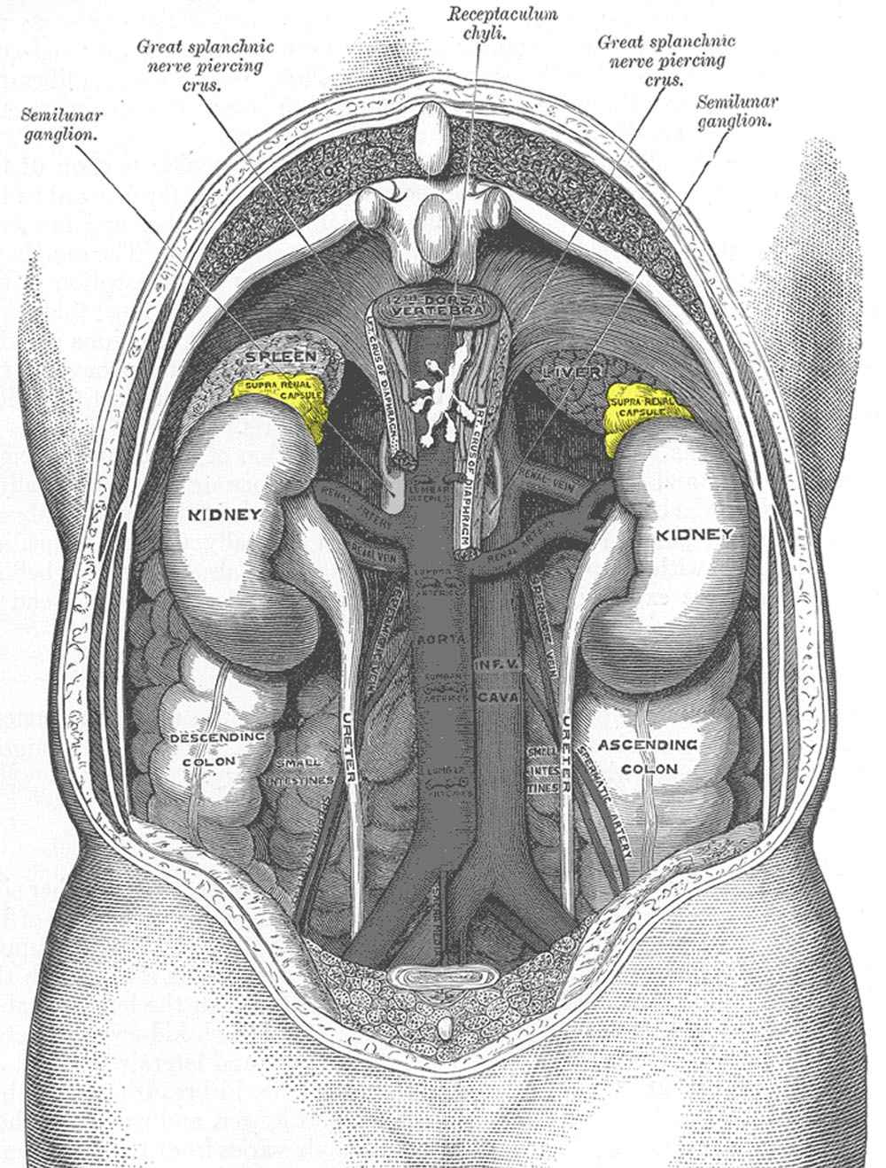
Roxbury-de Gray1120: adrenal glands in yellow.
16) Endothelial dysfunction drives oxidative stress and inflammation in Long COVID:
Both endothelial dysfunction and oxidative stress play an important part in the pathophysiology of Long COVID. Your blood vessels and lungs are lined with endothelial cells. Endothelial dysfunction can alter cell permeability and cause fluid build up which increases blood pressure (Izzo et al. 2022).
Chronic stress and anxiety can influence all aspects of the cardiovascular system. Reducing stress can lower blood pressure in many people.
Want more Long COVID causes? Check out how unhealthy mitochondria might be causing Long COVID.
Did you miss the first thrilling installment in What Causes Long COVID? Immune System Glitches.
Part 1 includes six exciting reasons your immune system may be doing you dirty:
- 1) Unwanted house guest. The virus is still present and causing problem.
- 2) Your immune cells are quietly quitting.
- 3) Zombie invasion! Virus infection summons zombie viruses already laying latent in your body.
- 4) Instead of quitting; your immune system went full on crazy after COVID infection and started packing heat.
- 5) Self sabotage. You start making autoantibodies that attack yourself.
- 6) Your immune system is dialed up too high: The interferon pathway story.
If all these Long COVID causes are getting depressing, here is a picture of a baby goat to brighten your day.

Definitions:
Brain-gut axis is based on how the brain and the gut communication with each other. This complex network involves crosstalk between the vagus nerve; the central nervous system; and gut microbiota; using neural, endocrine, immune, and humoral links. It regulates gastrointestinal homeostasis and it connects the emotional and cognitive areas of the brain with gut functions.
Cytokine storms are life-threatening systemic inflammatory syndromes which involve dangerously high levels of circulating inflammatory chemicals, cytokines, along with hyperactivation of the immune system.
Post-exertional malaise (PEM) which is also called post-exertional symptom exacerbation (PESE) or post-exertional neuroimmune exhaustion(PENE): a medical condition that causes symptoms to get worse after even minor physical or mental exertion. It is common in Long COVID, fibromyalgia, and myalgic encephalomyelitis or chronic fatigue syndrome (ME/CFS).
By Susan Fluegel PHD Nutritional Biochemistry
* Names and some other small details have been changed to protect people's privacy.
This information is for informational purposes only and does not constitute medical advice, diagnosis, or treatment.
References:
Banu H, Sultana N, Md Shahed M, Hasanat MA, Saleh AA, Arafat SM. Hypothalamic-Pituitary-Adrenal Axis Activity in SARS-CoV-2 Infected Noncritically Ill Hospitalized Patients. J ASEAN Fed Endocr Soc. 2023;38(2):65-70. doi: 10.15605/jafes.038.02.04. Full paper.
Breit S, Kupferberg A, Rogler G, Hasler G. Vagus Nerve as Modulator of the Brain-Gut Axis in Psychiatric and Inflammatory Disorders. Front Psychiatry. 2018 Mar 13;9:44. doi: 10.3389/fpsyt.2018.00044. Full article.
Buoite Stella A, Furlanis G, Frezza NA, Valentinotti R, Ajcevic M, Manganotti P. Autonomic dysfunction in post-COVID patients with and witfhout neurological symptoms: a prospective multidomain observational study. J Neurol. 2022 Feb;269(2):587-596. doi: 10.1007/s00415-021-10735-y. Full article.
Cervia-Hasler C, Brüningk SC, Hoch T, Fan B, Muzio G, Thompson RC, Ceglarek L, Meledin R, Westermann P, Emmenegger M, Taeschler P, Zurbuchen Y, Pons M, Menges D, Ballouz T, Cervia-Hasler S, Adamo S, Merad M, Charney AW, Puhan M, Brodin P, Nilsson J, Aguzzi A, Raeber ME, Messner CB, Beckmann ND, Borgwardt K, Boyman O. Persistent complement dysregulation with signs of thromboinflammation in active Long Covid. Science. 2024 Jan 19;383(6680):eadg7942. doi: 10.1126/science.adg7942. Epub 2024 Jan 19. Full article.
Collete M, Dos Santos TR, de Araújo N, Martins APC, Nagashima S, Vaz de Paula CB, Machado-Souza C, de Noronha L. Neutrophil Extracellular Trap Markers in Post Mortem Lung Biopsies from COVID-19 Patients. Int J Mol Sci. 2025 Aug 20;26(16):8059. doi: 10.3390/ijms26168059. Full article.
Crow MK, Ronnblom L. Type I interferons in host defence and inflammatory diseases. Lupus Sci Med. 2019 May 28;6(1):e000336. doi: 10.1136/lupus-2019-000336. Full article.
Dai Y, Zhou J, Shi C. Inflammasome: structure, biological functions, and therapeutic targets. MedComm (2020). 2023 Oct 9;4(5):e391. doi: 10.1002/mco2.391. Full article.
de Boer E, Petrache I, Goldstein NM, Olin JT, Keith RC, Modena B, Mohning MP, Yunt ZX, San-Millán I, Swigris JJ. Decreased Fatty Acid Oxidation and Altered Lactate Production during Exercise in Patients with Post-acute COVID-19 Syndrome. Am J Respir Crit Care Med. 2022 Jan 1;205(1):126-129. doi: 10.1164/rccm.202108-1903LE. Full article.
Davis HE, McCorkell L, Vogel JM, Topol EJ. Long COVID: major findings, mechanisms and recommendations. Nat Rev Microbiol. 2023 Mar;21(3):133-146. doi: 10.1038/s41579-022-00846-2. Epub 2023 Jan 13. Erratum in: Nat Rev Microbiol. 2023 Jun;21(6):408. doi: 10.1038/s41579-023-00896-0. Full article.
Durcan E, Hacioglu A, Karaca Z, Unluhizarci K, Gonen MS, Kelestimur F. Hypothalamic-Pituitary Axis Function and Adrenal Insufficiency in COVID-19 Patients. Neuroimmunomodulation. 2023;30(1):215-225. doi: 10.1159/000534025. Full article.
Excess weight, not high blood sugar, associated with increased risk of COVID-19 infection and long COVID. News release. EurekAlert. September 16, 2022. Accessed September 30, 2022. https://www.eurekalert.org/news-releases/964905
Fernández-Castañeda A, Lu P, Geraghty AC, Song E, Lee MH, Wood J, Yalçin B, Taylor KR, Dutton S, Acosta-Alvarez L, Ni L, Contreras-Esquivel D, Gehlhausen JR, Klein J, Lucas C, Mao T, Silva J, Peña-Hernández MA, Tabachnikova A, Takahashi T, Tabacof L, Tosto-Mancuso J, Breyman E, Kontorovich A, McCarthy D, Quezado M, Hefti M, Perl D, Folkerth R, Putrino D, Nath A, Iwasaki A, Monje M. Mild respiratory SARS-CoV-2 infection can cause multi-lineage cellular dysregulation and myelin loss in the brain. bioRxiv [Preprint]. 2022 Jan 10:2022.01.07.475453. doi: 10.1101/2022.01.07.475453. Full article.
Frontera JA, Sabadia S, Yang D, de Havenon A, Yaghi S, Lewis A, Lord AS, Melmed K, Thawani S, Balcer LJ, Wisniewski T, Galetta SL; NYU Neurology COVID-19 Study Team. Life stressors significantly impact long-term outcomes and post-acute symptoms 12-months after COVID-19 hospitalization. J Neurol Sci. 2022 Nov 5;443:120487. doi: 10.1016/j.jns.2022.120487. Full article.
Feng J, Li J, Li Y, Jin Y, Du F, Chen X. Elevated Serum D-Dimer May Reflect the Presence of Gut Inflammation in Spondyloarthritis. Front Med (Lausanne). 2022 Jan 21;8:816422. doi: 10.3389/fmed.2021.816422. Full article.
Genecand L, Altarelli M, Binkova A, Loew S, Vaudan S, Gex G, Bridevaux PO, Frésard I. Dysfunctional breathing symptoms, functional impact and quality of life in patients with long COVID-19: a prospective case series. BMJ Open Respir Res. 2023 Jul;10(1):e001770. doi: 10.1136/bmjresp-2023-001770. Full article.
Georgieva E, Ananiev J, Yovchev Y, Arabadzhiev G, Abrashev H, Abrasheva D, Atanasov V, Kostandieva R, Mitev M, Petkova-Parlapanska K, Karamalakova Y, Koleva-Korkelia I, Tsoneva V, Nikolova G. COVID-19 Complications: Oxidative Stress, Inflammation, and Mitochondrial and Endothelial Dysfunction. Int J Mol Sci. 2023 Oct 4;24(19):14876. doi: 10.3390/ijms241914876. Full article.
Giunta S, Giordani C, De Luca M, Olivieri F. Long-COVID-19 autonomic dysfunction: An integrated view in the framework of inflammaging. Mech Ageing Dev. 2024 Apr;218:111915. doi: 10.1016/j.mad.2024.111915. Summary.
Goh D, Lim JCT, Fernaíndez SB, Joseph CR, Edwards SG, Neo ZW, Lee JN, Caballero SG, Lau MC, Yeong JPS. Case report: Persistence of residual antigen and RNA of the SARS-CoV-2 virus in tissues of two patients with long COVID. Front Immunol. 2022 Sep 5;13:939989. doi: 10.3389/fimmu.2022.939989. Erratum in: Front Immunol. 2022 Oct 06;13:1036894. Full article.
Gupta K, Toelzer C, Williamson MK, Shoemark DK, Oliveira ASF, Matthews DA, Almuqrin A, Staufer O, Yadav SKN, Borucu U, Garzoni F, Fitzgerald D, Spatz J, Mulholland AJ, Davidson AD, Schaffitzel C, Berger I. Structural insights in cell-type specific evolution of intra-host diversity by SARS-CoV-2. Nat Commun. 2022 Jan 11;13(1):222. doi: 10.1038/s41467-021-27881-6.Full article.
Greene C, Connolly R, Brennan D, Laffan A, O'Keeffe E, Zaporojan L, O'Callaghan J, Thomson B, Connolly E, Argue R, Meaney JFM, Martin-Loeches I, Long A, Cheallaigh CN, Conlon N, Doherty CP, Campbell M. Blood-brain barrier disruption and sustained systemic inflammation in individuals with long COVID-associated cognitive impairment. Nat Neurosci. 2024 Mar;27(3):421-432. doi: 10.1038/s41593-024-01576-9. Epub 2024 Feb 22. Erratum in: Nat Neurosci. 2024 May;27(5):1019. doi: 10.1038/s41593-024-01644-0. Full article.
Heerdt PM, Shelley B, Singh I. Impaired systemic oxygen extraction long after mild COVID-19: potential perioperative implications. Br J Anaesth. 2022 Mar;128(3):e246-e249. doi: 10.1016/j.bja.2021.12.036. Epub 2021 Dec 27. Erratum in: Br J Anaesth. 2022 Nov;129(5):829-830. Full article.
Izzo R, Trimarco V, Mone P, Aloè T, Capra Marzani M, Diana A, Fazio G, Mallardo M, Maniscalco M, Marazzi G, Messina N, Mininni S, Mussi C, Pelaia G, Pennisi A, Santus P, Scarpelli F, Tursi F, Zanforlin A, Santulli G, Trimarco B. Combining L-Arginine with vitamin C improves long-COVID symptoms: The LINCOLN Survey. Pharmacol Res. 2022 Sep;183:106360. doi: 10.1016/j.phrs.2022.106360. Full article.
Kanczkowski W, Beuschlein F, & Bornstein, SR. Is there a role for the adrenal glands in long COVID?. Nat Rev Endocrinol 18, 451–452 (2022). https://doi.org/10.1038/s41574-022-00700-8 Full article.
Kell DB, Laubscher GJ, Pretorius E. A central role for amyloid fibrin microclots in long COVID/PASC: origins and therapeutic implications. Biochem J. 2022 Feb 17;479(4):537-559. doi: 10.1042/BCJ20220016. Full article.
Klein J, Wood J, Jaycox J, Lu P, Dhodapkar RM, Gehlhausen JR, Tabachnikova A, Tabacof L, Malik AA, Kamath K, Greene K, Monteiro VS, Peña-Hernandez M, Mao T, Bhattacharjee B, Takahashi T, Lucas C, Silva J, Mccarthy D, Breyman E, Tosto-Mancuso J, Dai Y, Perotti E, Akduman K, Tzeng TJ, Xu L, Yildirim I, Krumholz HM, Shon J, Medzhitov R, Omer SB, van Dijk D, Ring AM, Putrino D, Iwasaki A. Distinguishing features of Long COVID identified through immune profiling. medRxiv [Preprint]. 2022 Aug 10:2022.08.09.22278592. doi: 10.1101/2022.08.09.22278592. Full article.
Koch CA. Long Covid: Hormone Imbalances and/or Rather Complex Immune Dysregulations? J Endocr Soc. 2024 Mar 6;8(5):bvae043. doi: 10.1210/jendso/bvae043. Full article.
Komaroff AL, Lipkin WI. ME/CFS and Long COVID share similar symptoms and biological abnormalities: road map to the literature. Front Med (Lausanne). 2023 Jun 2;10:1187163. doi: 10.3389/fmed.2023.1187163. Full article.
Larsen, Nicholas & Stiles, Lauren & Shaik, Ruba & Schneider, Logan & Muppidi, Srikanth & Tsui, Cheuk & Geng, Linda & Bonilla, Hector & Miglis, Mitchell. (2022). Characterization of autonomic symptom burden in long COVID: A global survey of 2,314 adults. Frontiers in Neurology. 13. 10.3389/fneur.2022.1012668. Full article.
Liu Y, Ebinger JE, Mostafa R, Budde P, Gajewski J, Walker B, Joung S, Wu M, Bräutigam M, Hesping F, Rupieper E, Schubert AS, Zucht HD, Braun J, Melmed GY, Sobhani K, Arditi M, Van Eyk JE, Cheng S, Fert-Bober J. Paradoxical sex-specific patterns of autoantibody response to SARS-CoV-2 infection. J Transl Med. 2021 Dec 30;19(1):524. doi: 10.1186/s12967-021-03184-8. Full article.
Liu Q, Mak JWY, Su Q, Yeoh YK, Lui GC, Ng SSS, Zhang F, Li AYL, Lu W, Hui DS, Chan PK, Chan FKL, Ng SC. Gut microbiota dynamics in a prospective cohort of patients with post-acute COVID-19 syndrome. Gut. 2022 Mar;71(3):544-552. doi: 10.1136/gutjnl-2021-325989. Full article.
Madsen HB, Durhuus JA, Andersen O, thor Staten P, Rahbech A, Desler D. Mitochondrial dysfunction in acute and post-acute phases of COVID-19 and risk of non-communicable diseases. npj Metab Health Dis 2, 36 (2024). https://doi.org/10.1038/s44324-024-00038-x Full article.
Martínez-Colón GJ, Ratnasiri K, Chen H, Jiang S, Zanley E, Rustagi A, Verma R, Chen H, Andrews JR, Mertz KD, Tzankov A, Azagury D, Boyd J, Nolan GP, Schürch CM, Matter MS, Blish CA, McLaughlin TL. SARS-CoV-2 infection drives an inflammatory response in human adipose tissue through infection of adipocytes and macrophages. Sci Transl Med. 2022 Sep 22:eabm9151. doi: 10.1126/scitranslmed.abm9151. Full article.
Molnar T, Lehoczki A, Fekete M, Varnai R, Zavori L, Erdo-Bonyar S, Simon D, Berki T, Csecsei P, Ezer E. Mitochondrial dysfunction in long COVID: mechanisms, consequences, and potential therapeutic approaches. Geroscience. 2024 Apr 26. doi: 10.1007/s11357-024-01165-5. Full article.
Motiejunaite J, Balagny P, Arnoult F, Mangin L, Bancal C, d'Ortho MP, Frija-Masson J. Hyperventilation: A Possible Explanation for Long-Lasting Exercise Intolerance in Mild COVID-19 Survivors? Front Physiol. 2021 Jan 18;11:614590. doi: 10.3389/fphys.2020.614590. Full article.
Natarajan A, Zlitni S, Brooks EF, Vance SE, Dahlen A, Hedlin H, Park RM, Han A, Schmidtke DT, Verma R, Jacobson KB, Parsonnet J, Bonilla HF, Singh U, Pinsky BA, Andrews JR, Jagannathan P, Bhatt AS. Gastrointestinal symptoms and fecal shedding of SARS-CoV-2 RNA suggest prolonged gastrointestinal infection. Med (N Y). 2022 Jun 10;3(6):371-387.e9. doi: 10.1016/j.medj.2022.04.001. Full article.
Norweg A, Yao L, Barbuto S, Nordvig AS, Tarpey T, Collins E, Whiteson J, Sweeney G, Haas F, Leddy J. Exercise intolerance associated with impaired oxygen extraction in patients with long COVID. Respir Physiol Neurobiol. 2023 Jul;313:104062. doi: 10.1016/j.resp.2023.104062. Full article.
Pasquesi GIM, Allen H, Ivancevic A, Barbachano-Guerrero A, Joyner O, Guo K, Simpson DM, Gapin K, Horton I, Nguyen L, Yang Q, Warren CJ, Florea LD, Bitler BG, Santiago ML, Sawyer SL, Chuong EB. Regulation of human interferon signaling by transposon exonization. bioRxiv [Preprint]. 2023 Sep 15:2023.09.11.557241. doi: 10.1101/2023.09.11.557241. Full article.
Peker BO, Sener AG, Kaptan Aydogmus F. Antinuclear antibodies (ANAs) detected by indirect immunofluorescence (IIF) method in acute COVID-19 infection; future roadmap for laboratory diagnosis. J Immunol Methods. 2021 Dec;499:113174. doi: 10.1016/j.jim.2021.113174. Full article.
Phetsouphanh C, Darley DR, Wilson DB, Howe A, Munier CML, Patel SK, Juno JA, Burrell LM, Kent SJ, Dore GJ, Kelleher AD, Matthews GV. Immunological dysfunction persists for 8 months following initial mild-to-moderate SARS-CoV-2 infection. Nat Immunol. 2022 Feb;23(2):210-216. doi: 10.1038/s41590-021-01113-x. Full article.
Phetsouphanh C, Jacka B, Ballouz S, Jackson KJL, Wilson DB, Manandhar B, Klemm V, Tan HX, Wheatley A, Aggarwal A, Akerman A, Milogiannakis V, Starr M, Cunningham P, Turville SG, Kent SJ, Byrne A, Brew BJ, Darley DR, Dore GJ, Kelleher AD, Matthews GV. Improvement of immune dysregulation in individuals with long COVID at 24-months following SARS-CoV-2 infection. Nat Commun. 2024 Apr 17;15(1):3315. doi: 10.1038/s41467-024-47720-8. Full article.
Prasannan N, Heightman M, Hillman T, Wall E, Bell R, Kessler A, Neave L, Doyle A, Devaraj A, Singh D, Dehbi HM, Scully M. Impaired exercise capacity in post-COVID-19 syndrome: the role of VWF-ADAMTS13 axis. Blood Adv. 2022 Jul 12;6(13):4041-4048. doi: 10.1182/bloodadvances.2021006944. Full article.
Proal AD, VanElzakker MB. Long COVID or Post-acute Sequelae of COVID-19 (PASC): An Overview of Biological Factors That May Contribute to Persistent Symptoms. Front Microbiol. 2021 Jun 23;12:698169. doi: 10.3389/fmicb.2021.698169. Full article.
Ryu JK, Yan Z, Montano M, Sozmen EG, Dixit K, Suryawanshi RK, Matsui Y, Helmy E, Kaushal P, Makanani SK, Deerinck TJ, Meyer-Franke A, Rios Coronado PE, Trevino TN, Shin MG, Tognatta R, Liu Y, Schuck R, Le L, Miyajima H, Mendiola AS, Arun N, Guo B, Taha TY, Agrawal A, MacDonald E, Aries O, Yan A, Weaver O, Petersen MA, Meza Acevedo R, Alzamora MDPS, Thomas R, Traglia M, Kouznetsova VL, Tsigelny IF, Pico AR, Red-Horse K, Ellisman MH, Krogan NJ, Bouhaddou M, Ott M, Greene WC, Akassoglou K. Fibrin drives thromboinflammation and neuropathology in COVID-19. Nature. 2024 Sep;633(8031):905-913. doi: 10.1038/s41586-024-07873-4. Full article.
Saito S, Shahbaz S, Luo X, Osman M, Redmond D, Cohen Tervaert JW, Li L, Elahi S. Metabolomic and immune alterations in long COVID patients with chronic fatigue syndrome. Front Immunol. 2024 Jan 18;15:1341843. doi: 10.3389/fimmu.2024.1341843. Full article.
Saleh J, Peyssonnaux C, Singh KK, Edeas M. Mitochondria and microbiota dysfunction in COVID-19 pathogenesis. Mitochondrion. 2020 Sep;54:1-7. doi: 10.1016/j.mito.2020.06.008. Full article.
Singh I, Joseph P, Heerdt PM, Cullinan M, Lutchmansingh DD, Gulati M, Possick JD, Systrom DM, Waxman AB. Persistent Exertional Intolerance After COVID-19: Insights From Invasive Cardiopulmonary Exercise Testing. Chest. 2022 Jan;161(1):54-63. doi: 10.1016/j.chest.2021.08.010. Full article.
Shouman K, Vanichkachorn G, Cheshire WP, Suarez MD, Shelly S, Lamotte GJ, Sandroni P, Benarroch EE, Berini SE, Cutsforth-Gregory JK, Coon EA, Mauermann ML, Low PA, Singer W. Autonomic dysfunction following COVID-19 infection: an early experience. Clin Auton Res. 2021 Jun;31(3):385-394. doi: 10.1007/s10286-021-00803-8. Full article.
Son K, Jamil R, Chowdhury A, Mukherjee M, Venegas C, Miyasaki K, Zhang K, Patel Z, Salter B, Yuen ACY, Lau KS, Cowbrough B, Radford K, Huang C, Kjarsgaard M, Dvorkin-Gheva A, Smith J, Li QZ, Waserman S, Ryerson CJ, Nair P, Ho T, Balakrishnan N, Nazy I, Bowdish DME, Svenningsen S, Carlsten C, Mukherjee M. Circulating anti-nuclear autoantibodies in COVID-19 survivors predict long COVID symptoms. Eur Respir J. 2023 Jan 12;61(1):2200970. doi: 10.1183/13993003.00970-2022. Full article.
Stefano GB, Büttiker P, Weissenberger S, Ptacek R, Wang F, Esch T, Bilfinger TV, Raboch J, Kream RM. Biomedical Perspectives of Acute and Chronic Neurological and Neuropsychiatric Sequelae of COVID-19. Curr Neuropharmacol. 2022;20(6):1229-1240. doi: 10.2174/1570159X20666211223130228. Full article.
Stein SR, Ramelli SC, Grazioli A, Chung JY, Singh M, Yinda CK, Winkler CW, Sun J, Dickey JM, Ylaya K, Ko SH, Platt AP, Burbelo PD, Quezado M, Pittaluga S, Purcell M, Munster VJ, Belinky F, Ramos-Benitez MJ, Boritz EA, Lach IA, Herr DL, Rabin J, Saharia KK, Madathil RJ, Tabatabai A, Soherwardi S, McCurdy MT; NIH COVID-19 Autopsy Consortium; Peterson KE, Cohen JI, de Wit E, Vannella KM, Hewitt SM, Kleiner DE, Chertow DS. SARS-CoV-2 infection and persistence in the human body and brain at autopsy. Nature. 2022 Dec;612(7941):758-763. doi: 10.1038/s41586-022-05542-y. Full article.
Su Y, Yuan D, Chen DG, Ng RH, Wang K, Choi J, Li S, Hong S, Zhang R, Xie J, Kornilov SA, Scherler K, Pavlovitch-Bedzyk AJ, Dong S, Lausted C, Lee I, Fallen S, Dai CL, Baloni P, Smith B, Duvvuri VR, Anderson KG, Li J, Yang F, Duncombe CJ, McCulloch DJ, Rostomily C, Troisch P, Zhou J, Mackay S, DeGottardi Q, May DH, Taniguchi R, Gittelman RM, Klinger M, Snyder TM, Roper R, Wojciechowska G, Murray K, Edmark R, Evans S, Jones L, Zhou Y, Rowen L, Liu R, Chour W, Algren HA, Berrington WR, Wallick JA, Cochran RA, Micikas ME; ISB-Swedish COVID-19 Biobanking Unit; Wrin T, Petropoulos CJ, Cole HR, Fischer TD, Wei W, Hoon DSB, Price ND, Subramanian N, Hill JA, Hadlock J, Magis AT, Ribas A, Lanier LL, Boyd SD, Bluestone JA, Chu H, Hood L, Gottardo R, Greenberg PD, Davis MM, Goldman JD, Heath JR. Multiple early factors anticipate post-acute COVID-19 sequelae. Cell. 2022 Mar 3;185(5):881-895.e20. doi: 10.1016/j.cell.2022.01.014. Full article.
Sureda A, Alizadeh J, Nabavi SF, Berindan-Neagoe I, Cismaru CA, Jeandet P, Los MJ, Clementi E, Nabavi SM, Ghavami S. Endoplasmic reticulum as a potential therapeutic target for covid-19 infection management? Eur J Pharmacol. 2020 Sep 5;882:173288. doi: 10.1016/j.ejphar.2020.173288. Full article.
Tarnawski AS, Ahluwalia A. Endothelial cells and blood vessels are major targets for COVID-19-induced tissue injury and spreading to various organs. World J Gastroenterol. 2022 Jan 21;28(3):275-289. doi: 10.3748/wjg.v28.i3.275. Full article.
Thierry AR, Usher T, Sanchez C, Turner S, Venter C, Pastor B, Waters M, Thompson A, Mirandola A, Pisareva E, Prevostel C, Laubscher GJ, Kell DB, Pretorius E. Circulating Microclots Are Structurally Associated With Neutrophil Extracellular Traps and Their Amounts Are Elevated in Long COVID Patients. J Med Virol. 2025 Oct;97(10):e70613. doi: 10.1002/jmv.70613. Full article.
Turner S, Khan MA, Putrino D, Woodcock A, Kell DB, Pretorius E. Long COVID: pathophysiological factors and abnormalities of coagulation. Trends Endocrinol Metab. 2023 Jun;34(6):321-344. doi: 10.1016/j.tem.2023.03.002. Epub 2023 Apr 19. Full article.
Wang S, Quan L, Chavarro JE, Slopen N, Kubzansky LD, Koenen KC, Kang JH, Weisskopf MG, Branch-Elliman W, Roberts AL. Associations of Depression, Anxiety, Worry, Perceived Stress, and Loneliness Prior to Infection With Risk of Post-COVID-19 Conditions. JAMA Psychiatry. 2022 Nov 1;79(11):1081-1091. doi: 10.1001/jamapsychiatry.2022.2640. Erratum in: JAMA Psychiatry. 2022 Sep 28;: Full article.
Wang M, Zhang Y, Li C, Chang W, Zhang L. The relationship between gut microbiota and COVID-19 progression: new insights into immunopathogenesis and treatment. Front Immunol. 2023 May 2;14:1180336. doi: 10.3389/fimmu.2023.1180336. Full article.
Wang B, Zhang L, Wang Y, Dai T, Qin Z, Zhou F, Zhang L. Alterations in microbiota of patients with COVID-19: potential mechanisms and therapeutic interventions. Signal Transduct Target Ther. 2022 Apr 29;7(1):143. doi: 10.1038/s41392-022-00986-0. Full article.
Wishart DS, Guo A, Oler E, Wang F, Anjum A, Peters H, Dizon R, Sayeeda Z, Tian S, Lee BL, Berjanskii M, Mah R, Yamamoto M, Jovel J, Torres-Calzada C, Hiebert-Giesbrecht M, Lui VW, Varshavi D, Varshavi D, Allen D, Arndt D, Khetarpal N, Sivakumaran A, Harford K, Sanford S, Yee K, Cao X, Budinski Z, Liigand J, Zhang L, Zheng J, Mandal R, Karu N, Dambrova M, Schiöth HB, Greiner R, Gautam V. HMDB 5.0: the Human Metabolome Database for 2022. Nucleic Acids Res. 2022 Jan 7;50(D1):D622-D631. doi: 10.1093/nar/gkab1062. Full article.
Wong AC, Devason AS, Umana IC, Cox TO, Dohnalová L, Litichevskiy L, Perla J, Lundgren P, Etwebi Z, Izzo LT, Kim J, Tetlak M, Descamps HC, Park SL, Wisser S, McKnight AD, Pardy RD, Kim J, Blank N, Patel S, Thum K, Mason S, Beltra JC, Michieletto MF, Ngiow SF, Miller BM, Liou MJ, Madhu B, Dmitrieva-Posocco O, Huber AS, Hewins P, Petucci C, Chu CP, Baraniecki-Zwil G, Giron LB, Baxter AE, Greenplate AR, Kearns C, Montone K, Litzky LA, Feldman M, Henao-Mejia J, Striepen B, Ramage H, Jurado KA, Wellen KE, O'Doherty U, Abdel-Mohsen M, Landay AL, Keshavarzian A, Henrich TJ, Deeks SG, Peluso MJ, Meyer NJ, Wherry EJ, Abramoff BA, Cherry S, Thaiss CA, Levy M. Serotonin reduction in post-acute sequelae of viral infection. Cell. 2023 Oct 26;186(22):4851-4867.e20. doi: 10.1016/j.cell.2023.09.013. Full article.
Viswanathan T, Misra A, Chan SH, Qi S, Dai N, Arya S, Martinez-Sobrido L, Gupta YK. A metal ion orients SARS-CoV-2 mRNA to ensure accurate 2'-O methylation of its first nucleotide. Nat Commun. 2021 Jun 2;12(1):3287. doi: 10.1038/s41467-021-23594-y. Full article.
Wang C, Chen L, Chen Y, Jia W, Cai X, Liu Y, Ji F, Xiong P, Liang A, Liu R, Guan Y, Cheng Z, Weng Y, Wang W, Duan Y, Kuang D, Xu S, Cai H, Xia Q, Yang D, Wang MW, Yang X, Zhang J, Cheng C, Liu L, Liu Z, Liang R, Wang G, Li Z, Xia H, Xia T. Abnormal global alternative RNA splicing in COVID-19 patients. PLoS Genet. 2022 Apr 14;18(4):e1010137. doi: 10.1371/journal.pgen.1010137. Full article.
Xu R, Lu R, Zhang T, Wu Q, Cai W, Han X, Wan Z, Jin X, Zhang Z, Zhang C. Temporal association between human upper respiratory and gut bacterial microbiomes during the course of COVID-19 in adults. Commun Biol. 2021 Feb 18;4(1):240. doi: 10.1038/s42003-021-01796-w. Full article.
Yin K, Peluso MJ, Luo X, Thomas R, Shin MG, Neidleman J, Andrew A, Young K, Ma T, Hoh R, Anglin K, Huang B, Argueta U, Lopez M, Valdivieso D, Asare K, Deveau TM, Munter SE, Ibrahim R, Ständker L, Lu S, Goldberg SA, Lee SA, Lynch KL, Kelly JD, Martin JN, Münch J, Deeks SG, Henrich TJ, Roan NR. Long COVID manifests with T cell dysregulation, inflammation, and an uncoordinated adaptive immune response to SARS-CoV-2. bioRxiv [Preprint]. 2023 Aug 4:2023.02.09.527892. doi: 10.1101/2023.02.09.527892. Update in: Nat Immunol. 2024 Feb;25(2):218-225. doi: 10.1038/s41590-023-01724-6. Full article.