The Wonderful Shades of Fat Cells
The Wonderful Shades of Fat Cells
Fat cells, known as adipocytes, come in five colors:
- ★ White
- ★ Brown
- ★ Beige
- ★ Pink
- ★ Yellow
Based on morphology (structure and function), there are five major adipose tissue types: white, pink, beige, brown, and yellow adipose tissue (Corrêa et al. 2019). Groups of fat cells are called adipose tissue.
Each of these fat cell shades has a different role to play
White adipocytes (WAT):
White fat cells get all the hate and none of the love. Nobody ever asks you to admire their fat gains or shows off their love handles. You need these chunky cells though! They are an important part of your endocrine system.
Healthy fat cells produce beneficial adipokines:
★ Adiponectin normalizes glucose homeostasis. It has anti-inflammatory, antidiabetic, and antiatherogenic effects.
★ Neuregulin reduces liver glucose production and fat storage (lipogenesis). Moderates cell survival, proliferation, migration, and differentiation.
★ Adipsin has neuroprotective effects; increases cell survival; and is positively associated with longevity. It stimulates adipose cells to make and store triacylglycerol. It improves glucose tolerance.
★ FGF21 extends lifespan and improves dysfunctions associated with aging.
★ Omentin is anti-inflammatory and increases insulin sensitivity
Unhealthy fat cells produce harmful adipokines:
★ Leptin regulates energy and appetite. It is negatively associated with longevity.
★ Chemerin is involved in cell proliferation, differentiation, and energy metabolism. It is negatively associated with longevity.
★ Resistin increases inflammation. It is positively correlated with cellular senescence (death), cell aging, insulin resistance and obesity.
★ Fetuin A (FetA) is positively associated with inflammation and insulin resistance.
★ Visfatin is positively associated with obesity and cancer risk. May promote cancer cell proliferation, angiogenesis, metastasis and drug resistance.
★ RBP4 is positively associated with insulin resistance and obesity. It impairs mitochondrial fatty acid β-oxidation.
White fat cells act as a reservoir of energy for the body. They store excess energy in the form of triglycerides (TG). TGs are fatty acids. White adipocytes contain a larger central lipid droplet and very few mitochondria.
During times of fasting or less energy the surplus energy in adipose tissue is released to other tissues.
Interesting fact: When you accumulate fat, adipose cells increase in both size and number. It is not healthy for fat cells to get too large. Hypertrophic adipocytes (extra large fat cells) become metabolically impaired which is associated with insulin resistance, metabolic disorders and inflammation. See what happens to these extra big fat cells here.

Did you know that bees gain fat for winter?
Bees who gain weight for the winter are called fat or diutinus bees. They have enlarged fat bodies to store lipids, greater protein reserves, and lower juvenile hormone levels.
Cold weather and less food trigger fat gain in bees. The changes are due to a blood protein called vitellogenin (sounds like name a pharmaceutical company would make up). Vitellogenin is a phospholipoglycoprotein. As you may guess from the name; phospholipoglycoprotein has properties of sugar, fat and proteins.
Fat makes cytokines and hormones
Adipose tissue is not passive; it makes up the body's largest endocrine organ. Your fat produces an incredible amount of biologically active compounds used to regulate the whole body.
Adipose tissue churns out over 50 different types of cell signaling proteins (called adipokines), as well as steroid hormones, immune factors, microRNA, cytokines, and more. Fat also modulates immune cell production of both inflammatory and anti-inflammatory cytokines.
Adipokines can modulate metabolism, energy use, reward system, immunity, inflammation, appetite and eating behavior, cardiovascular function, mood, behavior, stress and many other physiological and cognitive processes (discussion Clemente-Suárez et al. 2023, Park and Shimokawa 2024).
Exercise can help prevent adipose tissue (fat) dysfunction.

Fat and your gut bacteria are frenemies
Adipose tissue has a complex relationship with your gut bacteria. Either one can influence the other. Gut microbiotia composition and activity can influence how much and what types of adipokines are produced by fat cells (Suriano et al. 2020). Likewise, the chemical produced by fat cells (adipokines) can influence gut bacteria populations and gut inflammation (discussion Clemente-Suárez et al. 2023).
Science Bite: Cliff notes on how mitochondria work
Mitochondria collect the protons (H+) stripped from food and shutter them into their inner membrane using the electron transport chain (ETC). The ETC pumps protons across the mitochondrial inner membrane using ETC complexes I, III, and IV. Electrons are pulled through the ETC complexes using a series of reduction-oxidation reactions.
The buildup of protons in a small area generates protonmotive force (PMF); energy stored as an electrochemical gradient of protons. Mitochondria are basically biological batteries. Like a battery, PMF energy can be used to create heat or energy (as ATP).
Another way to imagine this is think of the mitochondria inner membrane as a dam holding back protons. Protons are continually pumped into the reservoir. Like a dam, energy is released when water (protons) escape the dam wall (inner membrane). Mitochondria use PMF to create heat using uncoupling proteins and/or energy using ATPases.
The bottom of ATPases are like small water wheels embedded into the mitochondria membrane. Protons trying to escape the crowding slip into the wheel and cause it to turn. The mechanical action of the ATPase turning rotates the flowerlike top. As the top turns it squeezes a phosphate (P) onto a ADP to make ATP. This mechanical action transfers energy into the high energy P-P bond. Later, your body can break the P-P bond and use the energy.
Brown adipocytes:
Brown fat cells are a key area for thermogenesis (creation of heat). They are also considered the holy grail of weight loss because they can burn a lot of calories. The brown color in the adipose tissue comes from greater concentration of blood vessels as well as the fat cells' high concentration of mitochondria (which contain vitamins B2, B6, B12, iron and energy transporting FADx).
Compared to white fat cells, brown fat cells are leaner, fitter and more energetic. They have denser mitochondria, more blood vessels, and smaller and more numerous fat deposits. Adults have around 25–100 g (0.9–3.5 oz) of brown adipose tissue.
Brown adipose cells express high levels of UCP1 (uncoupling protein-1) also cleverly called thermogenin. This mitochondrial inner membrane protein is used to turn on heat production. UCP1 opens up a hole in the mitochondria membrane that lets protons leak out. As they rush out, the excess energy is released as heat. This also dissipates the electrochemical proton gradient that is used to create ATP production by ATP synthase (discussion Crichton et al. 2017, Takeda et al. 2023, Jones et al. 2024).
Brown adipose tissue (BAT) reduces obesity-associated metabolic diseases and some cancers in humans (Takeda et al. 2023).
How brown fat is activated:
★ In adults, extended cold exposure, temperatures below 16°C (60.8°F), can activate brown fat and cause it to start producing extra heat.
★Cold exposure increases brown fat glucose uptake
★ Neuregulin stimulates thermogenesis in brown adipose tissue.
★ Discussion Peres Valgas da Silva et al. 2019
Mitochondria in brown fat have wide fluctuations in their metabolic rate depending on the background temperature (Brunetta et al. 2024).
Science Bite: What turns on the heat in brown fat?
Brown fat metabolism is negatively regulated by inhibitory factor 1 (IF1). When temperatures decrease, levels of IF1 decline; this turns on heat production (thermogenesis) in brown fat.
How it works: IF1 inhibits ATP synthase hydrolytic activity; this is the ability to generate mitochondrial membrane potential (MMP) through ATP hydrolysis. Hydrolysis is using water to breakdown molecules into smaller pieces. Basically, it is ATP synthase in reverse; instead of making energy (ATP) using MMP, it builds up the MMP (Brunetta et al. 2024).
*Mitochondrial membrane potential (MMP): a charge separation across the inner mitochondrial membrane that is created by the electron transport chain. Basically, protons (H+) stripped from food are ferried into the inner mitochondria membrane causing a buildup of positive hydrogen ions.
Brown fat improves metabolism:
★ Removes excess glucose and triglycerides from the bloodstream
★ Improves whole-body glucose disposal and insulin sensitivity
★ Improves glucose homeostasis
★ Stimulates lipolysis while increases uptake of circulating fatty acids and lipoproteins
★ Uses branched chain amino acids (BCAAs) for fuel
★ Activated brown fat produces adipokines (batokines) which can improve metabolic health. This includes fibroblast growth factor 21 (FGF21) which regulates lipid and glucose metabolism and lipokine 12,13-diHOME which increases fatty acid uptake in muscles and brown fat.
★ Discussion Peres Valgas da Silva et al. 2019, Jones et al. 2024
Beige/Brite adipocytes:
Beige fat cells are anything but sad. These caramel colored balls originate from white adipocytes. When white fat cells turn beige it is called browning.
Beige or brite fat develops when white fat is exposed to cold, exercise, or fasting. Cold exposure that leads to beige fat formation occurs at about 3°C above the person's shivering point. This is usually 11°C (51°F) for women and 9°C (48°F) for men; exercise; or fasting. Conversion of white fat into heat producing adipocytes is mostly due to peroxisome proliferator-activated receptor (PPARγ) activation (Batrow et al. 2025).
Beige fat cells share many of their characteristics with brown fat cells. They have an increased capacity for fuel oxidation and thermogenesis due to their increased mitochondrial density, multilocular lipid droplets, and higher levels of UCP1. UCP1 is under tight metabolic control and does not produce heat unless it is activated (Li and Fromme 2022).
In spite of the similarities, beige fat is genetically different than brown fat. Beige adipocytes have a high level of plasticity. This means that they can easily transform from white fat cells to beige fat cells and back again when exposed to different conditions (Corrêa et al. 2019).
Isoda Koryūsai - Painting the Eyes on a Snow Rabbit c1780.

Pink adipocytes:
Regulation of lactation (milk production) may involve white, brown and pink fat. Adipose cells in the mammary gland produce over 350 adipokines which act as hormones/in a regulatory function for both mother and baby. In particular, the interplay between white and brown fat may work together to maintain nutritional and energetic homeostasis (discussion Colleluori et al. 2021, Cinti 2025).
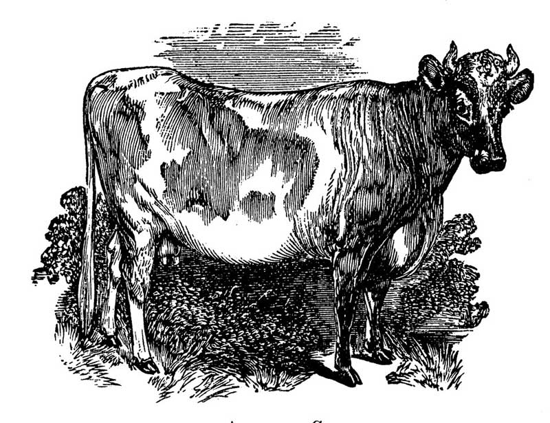
Alderney cow from Brett's Colonists' Guide c1883.
The mammary gland is an extremely plastic endocrine gland responsible for lactation. It is composed of some combination of adipose cells, epithelial cell,s and fibrous connective tissue; which varies depending on species. During pregnancy white adipocytes progressively seem to disappear leaving a space for newly formed milk producing alveoli. These get larger during lactation and transfer back into white adipose cells at the end of lactation. Some researchers call this in between stage during lactation pink fat cells.
Pink fat cells are formed from white adipocytes during pregnancy and lactation (milk production). This adipocyte epithelial cell combination contains milk containing granules. (Colleluori et al. 2021, Cinti 2025).
Yellow adipocytes:
Yellow fat cells are the marrow adipose tissues (MAT) in bones. Yellow adipocytes are anatomically and functionally separate from white, brown and beige fat cells. Their shape is similar to that of white fat cells, but they have different lipid profile, gene expression, and metabolism. Yellow marrow fat cells have a unilocular lipid store (one single fat deposit) and increases with age (Sebo et al. 2019, Pallio 2023).
Yellow bone marrow is a mix of fat cells and connective tissue. It stores fat and produces red blood cells (erythrocytes) and white blood cells (leukocytes). Like other adipose tissue, yellow fat cells produce adipokines. It is become clear that they regulate both bone and blood cell production and health. More research on this needs to be done, however.
By Susan Fluegel PHD Nutritional Biochemistry and Lori Wood MS Human Nutrition.
*Names and some minor identifying details in all stories in this website are changed to protect people's privacy.
I'm not your doctor so this is not medical information. I'm just a person who would like to see you happy and healthy. If you have any questions or concerns about starting an exercise regiment, diet program, or supplements please consult a professional.
References:
Batrow PL, Rooks CH, Gautier N, Martin L, Sibille B, Guillou H, Postic C, Langin D, Mothe-Satney I, Amri EZ. Regulation of UCP1 expression by PPARα and pemafibrate in human beige adipocytes. Life Sci. 2025 Feb 15;363:123406. doi: 10.1016/j.lfs.2025.123406. Abstract.
Brunetta HS, Jung AS, Valdivieso-Rivera F, de Campos Zani SC, Guerra J, Furino VO, Francisco A, Berçot M, Moraes-Vieira PM, Keipert S, Jastroch M, Martinez LO, Sponton CH, Castilho RF, Mori MA, Bartelt A. IF1 is a cold-regulated switch of ATP synthase hydrolytic activity to support thermogenesis in brown fat. EMBO J. 2024 Nov;43(21):4870-4891. doi: 10.1038/s44318-024-00215-0. Full article.
Cinti S. The triangle of transdifferentiation in the adipose organ, Obesity and Endocrinology, Volume 1, Issue 1, January 2025, wjaf005, https://doi.org/10.1093/obendo/wjaf005. Full article.
Clemente-Sárez VJ, Redondo-Flórez L, Beltrán-Velasco AI, Martín-Rodríguez A, Martínez-Guardado I, Navarro-Jiménez E, Laborde-Cárdenas CC, Tornero-Aguilera JF. The Role of Adipokines in Health and Disease. Biomedicines. 2023 Apr 27;11(5):1290. doi: 10.3390/biomedicines11051290. Full article.
Colleluori G, Perugini J, Barbatelli G, Cinti S. Mammary gland adipocytes in lactation cycle, obesity and breast cancer. Rev Endocr Metab Disord. 2021 Jun;22(2):241-255. doi: 10.1007/s11154-021-09633-5. Full article.
Corrêa LH, Heyn GS, Magalhaes KG. The Impact of the Adipose Organ Plasticity on Inflammation and Cancer Progression. Cells. 2019 Jun 30;8(7):662. doi: 10.3390/cells8070662. Full article.
Crichton PG, Lee Y, Kunji ER. The molecular features of uncoupling protein 1 support a conventional mitochondrial carrier-like mechanism. Biochimie. 2017 Mar;134:35-50. doi: 10.1016/j.biochi.2016.12.016. Full article.
Gómez-García I, Trepiana J, Fernández-Quintela A, Giralt M, Portillo MP. Sexual Dimorphism in Brown Adipose Tissue Activation and White Adipose Tissue Browning. Int J Mol Sci. 2022 Jul 26;23(15):8250. doi: 10.3390/ijms23158250. Full article.
Jones SA, Ruprecht JJ, Crichton PG, Kunji ERS. Structural mechanisms of mitochondrial uncoupling protein 1 regulation in thermogenesis. Trends Biochem Sci. 2024 Jun;49(6):506-519. doi: 10.1016/j.tibs.2024.03.005. Full article.
Li Y, Fromme T. Uncoupling Protein 1 Does Not Produce Heat without Activation. Int J Mol Sci. 2022 Feb 22;23(5):2406. doi: 10.3390/ijms23052406. Full article.
Pallio G. New Insights into Adipose Tissue Metabolic Function and Dysfunction. Int J Mol Sci. 2023 Jun 9;24(12):9953. doi: 10.3390/ijms24129953. Full article.
Park S, Shimokawa I. Influence of Adipokines on Metabolic Dysfunction and Aging. Biomedicines. 2024 Apr 15;12(4):873. doi: 10.3390/biomedicines12040873. Full article.
Peres Valgas da Silva C, Hernández-Saavedra D, White JD, Stanford KI. Cold and Exercise: Therapeutic Tools to Activate Brown Adipose Tissue and Combat Obesity. Biology (Basel). 2019 Feb 12;8(1):9. doi: 10.3390/biology8010009. Full article.
Senesi P, Luzi L, Terruzzi I. Adipokines, Myokines, and Cardiokines: The Role of Nutritional Interventions. Int J Mol Sci. 2020 Nov 8;21(21):8372. doi: 10.3390/ijms21218372. Full article.
Sebo ZL, Rendina-Ruedy E, Ables GP, Lindskog DM, Rodeheffer MS, Fazeli PK, Horowitz MC. Bone Marrow Adiposity: Basic and Clinical Implications. Endocr Rev. 2019 Oct 1;40(5):1187-1206. doi: 10.1210/er.2018-00138. Full article.
Suriano F, Van Hul M, Cani PD. Gut microbiota and regulation of myokine-adipokine function. Curr Opin Pharmacol. 2020 Jun;52:9-17. doi: 10.1016/j.coph.2020.03.006. Full article.
Takeda Y, Harada Y, Yoshikawa T, Dai P. Mitochondrial Energy Metabolism in the Regulation of Thermogenic Brown Fats and Human Metabolic Diseases. Int J Mol Sci. 2023 Jan 10;24(2):1352. doi: 10.3390/ijms24021352. Full article.